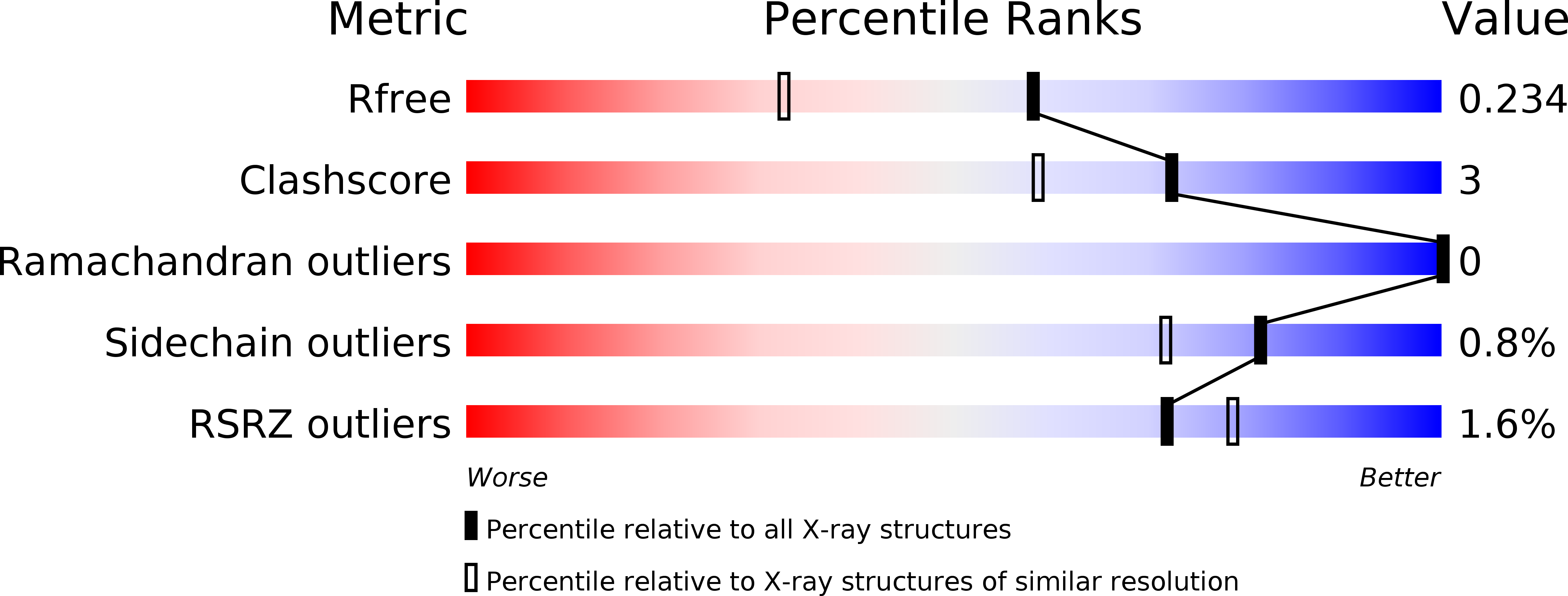
6Z3C image
Deposition Date 2020-05-19
Release Date 2020-06-03
Last Version Date 2024-01-24
Entry Detail
PDB ID:
6Z3C
Keywords:
Title:
High resolution structure of RgNanOx
Biological Source:
Source Organism:
Host Organism:
Method Details:
Experimental Method:
Resolution:
1.74 Å
R-Value Free:
0.22
R-Value Work:
0.18
Space Group:
P 21 21 21
Macromolecular Entities
Polymer Type:polypeptide(L)
Molecule:Gfo/Idh/MocA family oxidoreductase
Gene (Uniprot):CDL27_13940, DW270_01520, DW812_00100, DWY88_14550, DWZ50_08505, G4958_03345, G4993_04775, PNU93_07225, PNW85_04480
Chain IDs:A (auth: AAA), B (auth: BBB)
Chain Length:382
Number of Molecules:2
Biological Source:Ruminococcus gnavus
Primary Citation
Uncovering a novel molecular mechanism for scavenging sialic acids in bacteria.
J.Biol.Chem. 295 13724 13736 (2020)
PMID: 32669363 DOI: 10.1074/jbc.RA120.014454

Abstact

The human gut symbiont Ruminococcus gnavus scavenges host-derived N-acetylneuraminic acid (Neu5Ac) from mucins by converting it to 2,7-anhydro-Neu5Ac. We previously showed that 2,7-anhydro-Neu5Ac is transported into R. gnavus ATCC 29149 before being converted back to Neu5Ac for further metabolic processing. However, the molecular mechanism leading to the conversion of 2,7-anhydro-Neu5Ac to Neu5Ac remained elusive. Using 1D and 2D NMR, we elucidated the multistep enzymatic mechanism of the oxidoreductase (RgNanOx) that leads to the reversible conversion of 2,7-anhydro-Neu5Ac to Neu5Ac through formation of a 4-keto-2-deoxy-2,3-dehydro-N-acetylneuraminic acid intermediate and NAD+ regeneration. The crystal structure of RgNanOx in complex with the NAD+ cofactor showed a protein dimer with a Rossman fold. Guided by the RgNanOx structure, we identified catalytic residues by site-directed mutagenesis. Bioinformatics analyses revealed the presence of RgNanOx homologues across Gram-negative and Gram-positive bacterial species and co-occurrence with sialic acid transporters. We showed by electrospray ionization spray MS that the Escherichia coli homologue YjhC displayed activity against 2,7-anhydro-Neu5Ac and that E. coli could catabolize 2,7-anhydro-Neu5Ac. Differential scanning fluorimetry analyses confirmed the binding of YjhC to the substrates 2,7-anhydro-Neu5Ac and Neu5Ac, as well as to co-factors NAD and NADH. Finally, using E. coli mutants and complementation growth assays, we demonstrated that 2,7-anhydro-Neu5Ac catabolism in E. coli depended on YjhC and on the predicted sialic acid transporter YjhB. These results revealed the molecular mechanisms of 2,7-anhydro-Neu5Ac catabolism across bacterial species and a novel sialic acid transport and catabolism pathway in E. coli.

Legend

Protein

Chemical

Disease

Primary Citation of related structures