6Z23 image
Entry Detail
PDB ID:
6Z23
Title:
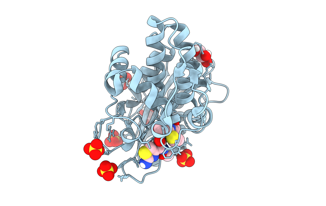
Acylenzyme complex of cefotaxime bound to deacylation mutant KPC-2 (E166Q)
Biological Source:
Source Organism:
PDB Version:
Deposition Date:
2020-05-14
Release Date:
2020-12-23
Method Details:
Experimental Method:
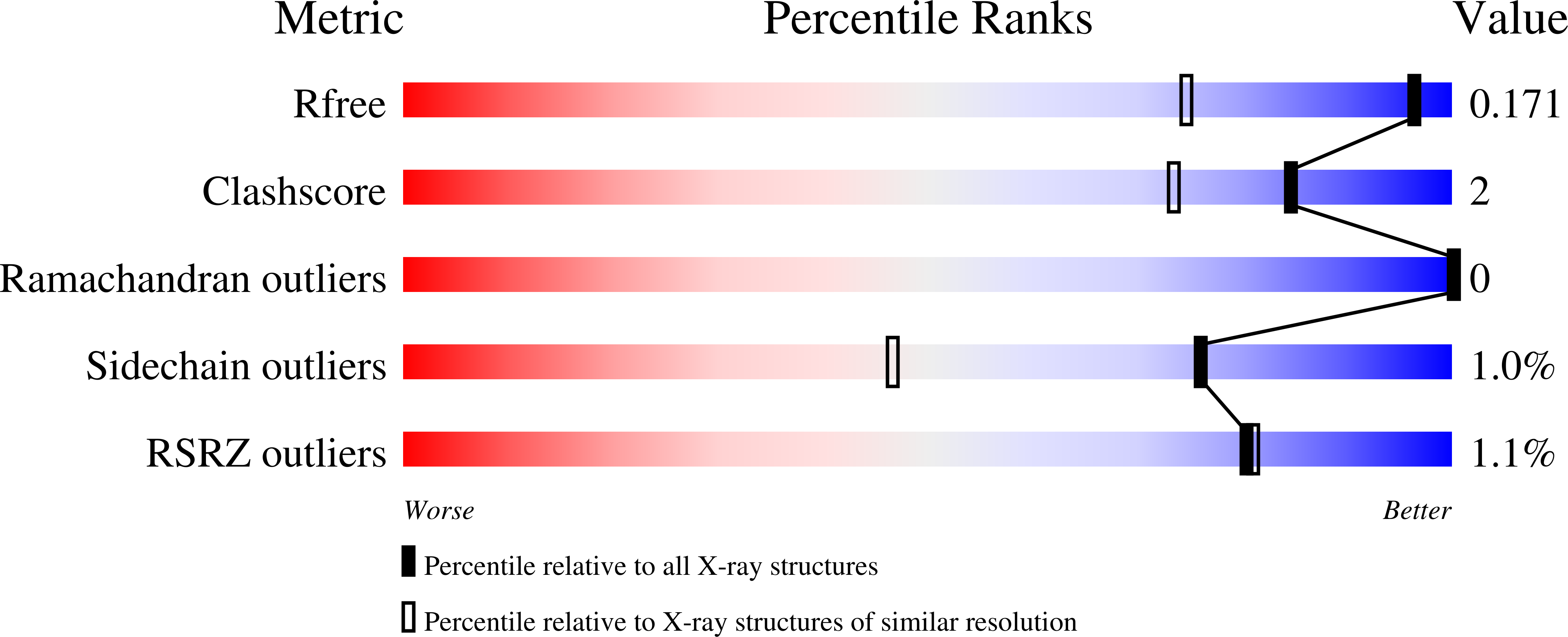
Resolution:
1.31 Å
R-Value Free:
0.17
R-Value Work:
0.14
R-Value Observed:
0.14
Space Group:
P 21 21 2
Macromolecular Entities
Polymer Type:polypeptide(L)
Description:Carbapenem-hydrolyzing beta-lactamase KPC
Chain IDs:A
Chain Length:290
Number of Molecules:1
Biological Source:Klebsiella pneumoniae
Primary Citation
Natural variants modify Klebsiella pneumoniae carbapenemase (KPC) acyl-enzyme conformational dynamics to extend antibiotic resistance.
J.Biol.Chem. 296 100126 100126 (2020)
PMID: 33257320 DOI: 10.1074/jbc.RA120.016461

Abstact

Class A serine β-lactamases (SBLs) are key antibiotic resistance determinants in Gram-negative bacteria. SBLs neutralize β-lactams via a hydrolytically labile covalent acyl-enzyme intermediate. Klebsiella pneumoniae carbapenemase (KPC) is a widespread SBL that hydrolyzes carbapenems, the most potent β-lactams; known KPC variants differ in turnover of expanded-spectrum oxyimino-cephalosporins (ESOCs), for example, cefotaxime and ceftazidime. Here, we compare ESOC hydrolysis by the parent enzyme KPC-2 and its clinically observed double variant (P104R/V240G) KPC-4. Kinetic analyses show that KPC-2 hydrolyzes cefotaxime more efficiently than the bulkier ceftazidime, with improved ESOC turnover by KPC-4 resulting from enhanced turnover (kcat), rather than altered KM values. High-resolution crystal structures of ESOC acyl-enzyme complexes with deacylation-deficient (E166Q) KPC-2 and KPC-4 mutants show that ceftazidime acylation causes rearrangement of three loops; the Ω, 240, and 270 loops, which border the active site. However, these rearrangements are less pronounced in the KPC-4 than the KPC-2 ceftazidime acyl-enzyme and are not observed in the KPC-2:cefotaxime acyl-enzyme. Molecular dynamics simulations of KPC:ceftazidime acyl-enyzmes reveal that the deacylation general base E166, located on the Ω loop, adopts two distinct conformations in KPC-2, either pointing "in" or "out" of the active site; with only the "in" form compatible with deacylation. The "out" conformation was not sampled in the KPC-4 acyl-enzyme, indicating that efficient ESOC breakdown is dependent upon the ordering and conformation of the KPC Ω loop. The results explain how point mutations expand the activity spectrum of the clinically important KPC SBLs to include ESOCs through their effects on the conformational dynamics of the acyl-enzyme intermediate.

Legend

Protein

Chemical

Disease

Primary Citation of related structures