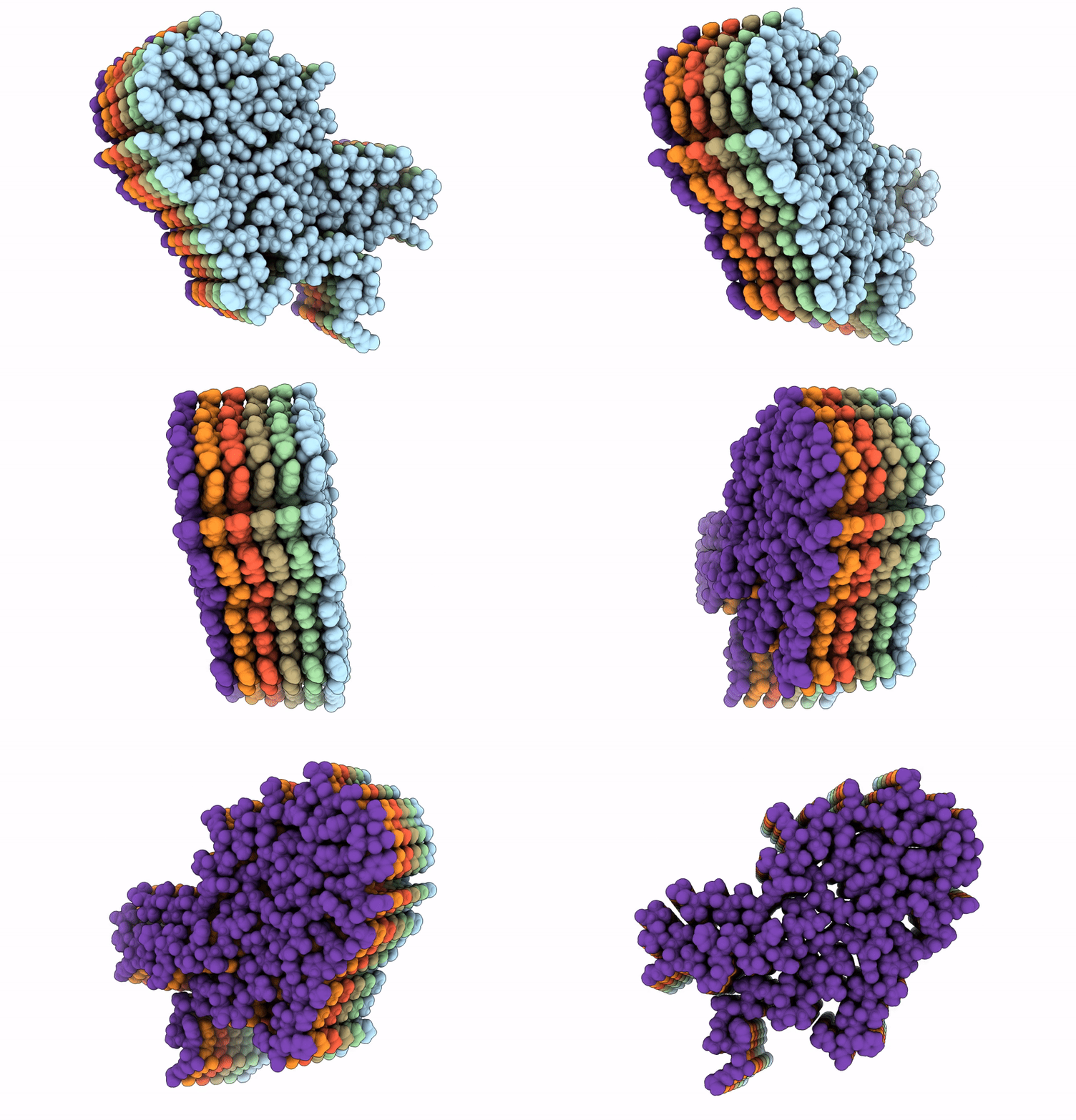
6Z1O image
Deposition Date 2020-05-14
Release Date 2021-02-24
Last Version Date 2024-10-09
Entry Detail
PDB ID:
6Z1O
Keywords:
Title:
AL amyloid fibril from a lambda 3 light chain in conformation A
Biological Source:
Source Organism:
Homo sapiens (Taxon ID: 9606)
Method Details:
Experimental Method:
Resolution:
3.20 Å
Aggregation State:
FILAMENT
Reconstruction Method:
HELICAL
Macromolecular Entities
Polymer Type:polypeptide(L)
Molecule:lambda 3 immunoglobulin light chain fragment, residues 2-116
Chain IDs:A (auth: D), B (auth: E), C (auth: F), D (auth: C), E (auth: B), F (auth: A)
Chain Length:89
Number of Molecules:6
Biological Source:Homo sapiens
Ligand Molecules
Primary Citation
Cryo-EM reveals structural breaks in a patient-derived amyloid fibril from systemic AL amyloidosis.
Nat Commun 12 875 875 (2021)
PMID: 33558536 DOI: 10.1038/s41467-021-21126-2

Abstact

Systemic AL amyloidosis is a debilitating and potentially fatal disease that arises from the misfolding and fibrillation of immunoglobulin light chains (LCs). The disease is patient-specific with essentially each patient possessing a unique LC sequence. In this study, we present two ex vivo fibril structures of a λ3 LC. The fibrils were extracted from the explanted heart of a patient (FOR005) and consist of 115-residue fibril proteins, mainly from the LC variable domain. The fibril structures imply that a 180° rotation around the disulfide bond and a major unfolding step are necessary for fibrils to form. The two fibril structures show highly similar fibril protein folds, differing in only a 12-residue segment. Remarkably, the two structures do not represent separate fibril morphologies, as they can co-exist at different z-axial positions within the same fibril. Our data imply the presence of structural breaks at the interface of the two structural forms.

Legend

Protein

Chemical

Disease

Primary Citation of related structures