6YSH image
Deposition Date 2020-04-22
Release Date 2020-09-02
Last Version Date 2024-01-24
Entry Detail
PDB ID:
6YSH
Keywords:
Title:
Lamin A 1-70 coil1A dimer stabilized by C-terminal capping
Biological Source:
Source Organism:
Homo sapiens (Taxon ID: 9606)
Host Organism:
Method Details:
Experimental Method:
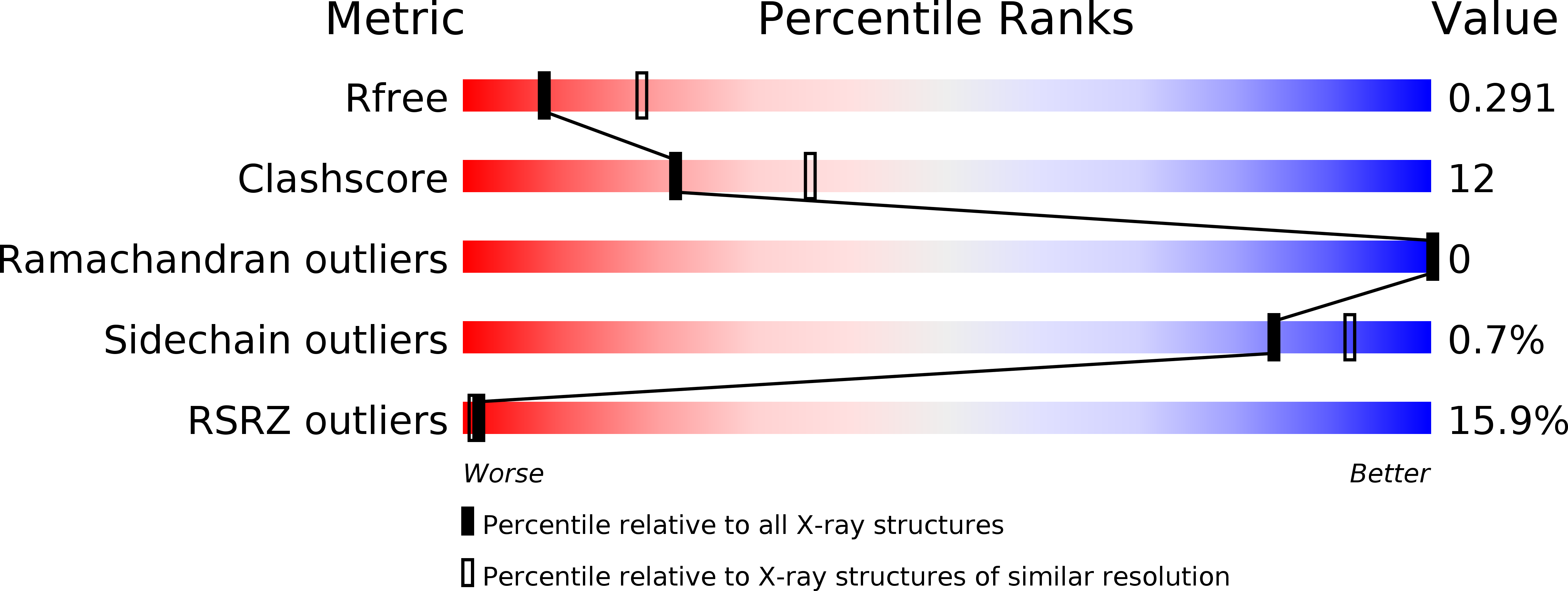
Resolution:
2.83 Å
R-Value Free:
0.28
R-Value Work:
0.23
R-Value Observed:
0.23
Space Group:
I 41 2 2
Macromolecular Entities
Protein Blast
Polymer Type:polypeptide(L)
Molecule:Prelamin-A/C,Microtubule-associated protein RP/EB family member 1
Gene (Uniprot):LMNA, MAPRE1
Chain IDs:A
Chain Length:83
Number of Molecules:1
Biological Source:Homo sapiens
Protein Blast
Polymer Type:polypeptide(L)
Molecule:Prelamin-A/C,Microtubule-associated protein RP/EB family member 1
Gene (Uniprot):LMNA, MAPRE1
Chain IDs:B
Chain Length:81
Number of Molecules:1
Biological Source:Homo sapiens
Primary Citation
Addressing the Molecular Mechanism of Longitudinal Lamin Assembly Using Chimeric Fusions.
Cells 9 ? ? (2020)
PMID: 32645958 DOI: 10.3390/cells9071633

Abstact

The molecular architecture and assembly mechanism of intermediate filaments have been enigmatic for decades. Among those, lamin filaments are of particular interest due to their universal role in cell nucleus and numerous disease-related mutations. Filament assembly is driven by specific interactions of the elementary dimers, which consist of the central coiled-coil rod domain flanked by non-helical head and tail domains. We aimed to investigate the longitudinal 'head-to-tail' interaction of lamin dimers (the so-called ACN interaction), which is crucial for filament assembly. To this end, we prepared a series of recombinant fragments of human lamin A centred around the N- and C-termini of the rod. The fragments were stabilized by fusions to heterologous capping motifs which provide for a correct formation of parallel, in-register coiled-coil dimers. As a result, we established crystal structures of two N-terminal fragments one of which highlights the propensity of the coiled-coil to open up, and one C-terminal rod fragment. Additional studies highlighted the capacity of such N- and C-terminal fragments to form specific complexes in solution, which were further characterized using chemical cross-linking. These data yielded a molecular model of the ACN complex which features a 6.5 nm overlap of the rod ends.

Legend

Protein

Chemical

Disease

Primary Citation of related structures
Feedback Form
Name
Email
Institute
Feedback