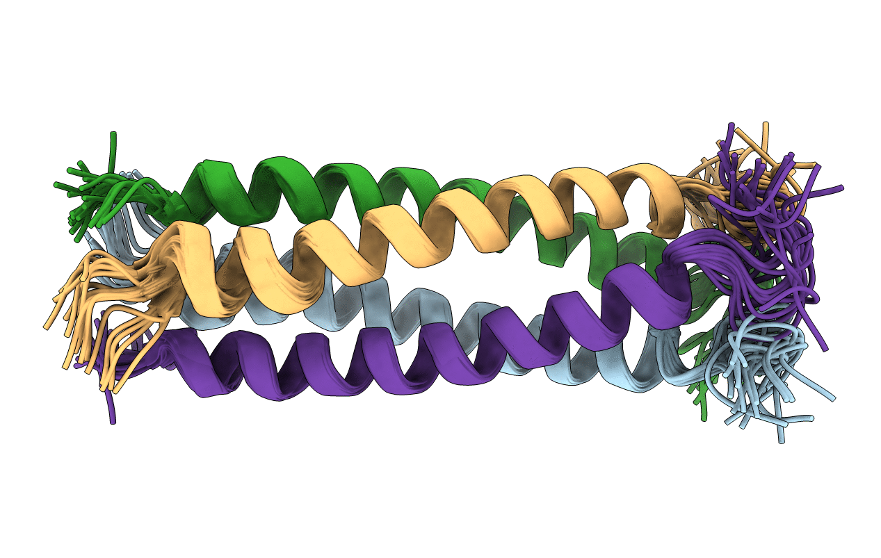
6YP5 image
Entry Detail
PDB ID:
6YP5
Keywords:
Title:
Solution NMR structure of the oligomerization domain of respiratory syncytial virus phosphoprotein
Biological Source:
PDB Version:
Deposition Date:
2020-04-15
Release Date:
2021-04-28
Method Details:
Experimental Method:
Conformers Calculated:
20
Conformers Submitted:
20
Selection Criteria:
all calculated structures submitted
Macromolecular Entities
Polymer Type:polypeptide(L)
Description:Phosphoprotein
Chain IDs:A, B, C, D
Chain Length:45
Number of Molecules:4
Biological Source:Respiratory syncytial virus
Ligand Molecules
Primary Citation
A Structural and Dynamic Analysis of the Partially Disordered Polymerase-Binding Domain in RSV Phosphoprotein.
Biomolecules 11 ? ? (2021)
PMID: 34439894 DOI: 10.3390/biom11081225

Abstact

The phosphoprotein P of Mononegavirales (MNV) is an essential co-factor of the viral RNA polymerase L. Its prime function is to recruit L to the ribonucleocapsid composed of the viral genome encapsidated by the nucleoprotein N. MNV phosphoproteins often contain a high degree of disorder. In Pneumoviridae phosphoproteins, the only domain with well-defined structure is a small oligomerization domain (POD). We previously characterized the differential disorder in respiratory syncytial virus (RSV) phosphoprotein by NMR. We showed that outside of RSV POD, the intrinsically disordered N-and C-terminal regions displayed a structural and dynamic diversity ranging from random coil to high helical propensity. Here we provide additional insight into the dynamic behavior of PCα, a domain that is C-terminal to POD and constitutes the RSV L-binding region together with POD. By using small phosphoprotein fragments centered on or adjacent to POD, we obtained a structural picture of the POD-PCα region in solution, at the single residue level by NMR and at lower resolution by complementary biophysical methods. We probed POD-PCα inter-domain contacts and showed that small molecules were able to modify the dynamics of PCα. These structural properties are fundamental to the peculiar binding mode of RSV phosphoprotein to L, where each of the four protomers binds to L in a different way.

Legend

Protein

Chemical

Disease

Primary Citation of related structures