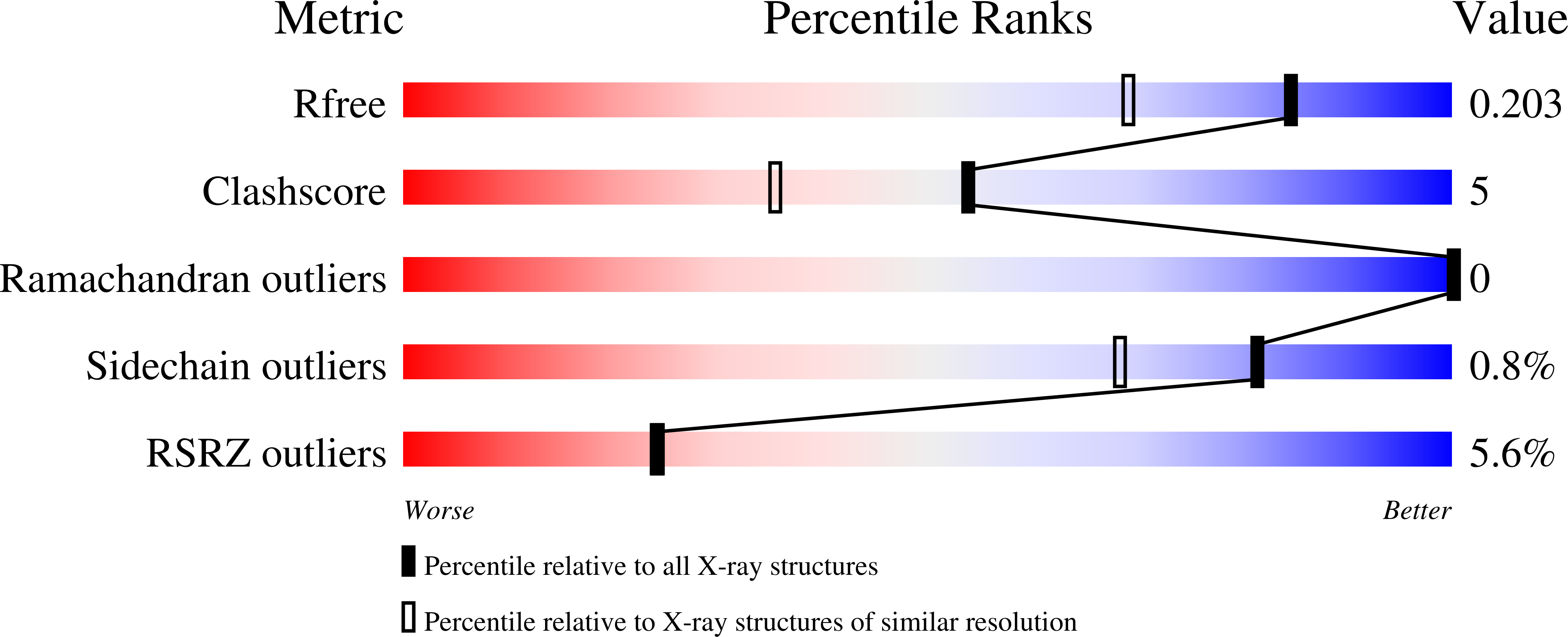
6YMT image
Deposition Date 2020-04-09
Release Date 2021-07-21
Last Version Date 2024-11-06
Entry Detail
PDB ID:
6YMT
Keywords:
Title:
RNASE 3/1 version1
Biological Source:
Source Organism:
Host Organism:
Method Details:
Experimental Method:
Resolution:
1.58 Å
R-Value Free:
0.20
R-Value Work:
0.17
R-Value Observed:
0.17
Space Group:
C 2 2 21
Macromolecular Entities
Polymer Type:polypeptide(L)
Molecule:RNase 3/1 version 1
Chain IDs:A (auth: G), B (auth: J), C, D, E, F, G (auth: A), H, I, J (auth: B), K (auth: L), L (auth: K)
Chain Length:128
Number of Molecules:12
Biological Source:synthetic construct
Ligand Molecules
Primary Citation
Exploring the RNase A scaffold to combine catalytic and antimicrobial activities. Structural characterization of RNase 3/1 chimeras.
Front Mol Biosci 9 964717 964717 (2022)
PMID: 36188223 DOI: 10.3389/fmolb.2022.964717

Abstact

Design of novel antibiotics to fight antimicrobial resistance is one of the first global health priorities. Novel protein-based strategies come out as alternative therapies. Based on the structure-function knowledge of the RNase A superfamily we have engineered a chimera that combines RNase 1 highest catalytic activity with RNase 3 unique antipathogen properties. A first construct (RNase 3/1-v1) was successfully designed with a catalytic activity 40-fold higher than RNase 3, but alas in detriment of its anti-pathogenic activity. Next, two new versions of the original chimeric protein were created showing improvement in the antimicrobial activity. Both second generation versions (RNases 3/1-v2 and -v3) incorporated a loop characteristic of RNase 3 (L7), associated to antimicrobial activity. Last, removal of an RNase 1 flexible loop (L1) in the third version enhanced its antimicrobial properties and catalytic efficiency. Here we solved the 3D structures of the three chimeras at atomic resolution by X-ray crystallography. Structural analysis outlined the key functional regions. Prediction by molecular docking of the protein chimera in complex with dinucleotides highlighted the contribution of the C-terminal region to shape the substrate binding cavity and determine the base selectivity and catalytic efficiency. Nonetheless, the structures that incorporated the key features related to RNase 3 antimicrobial activity retained the overall RNase 1 active site conformation together with the essential structural elements for binding to the human ribonuclease inhibitor (RNHI), ensuring non-cytotoxicity. Results will guide us in the design of the best RNase pharmacophore for anti-infective therapies.

Legend

Protein

Chemical

Disease

Primary Citation of related structures