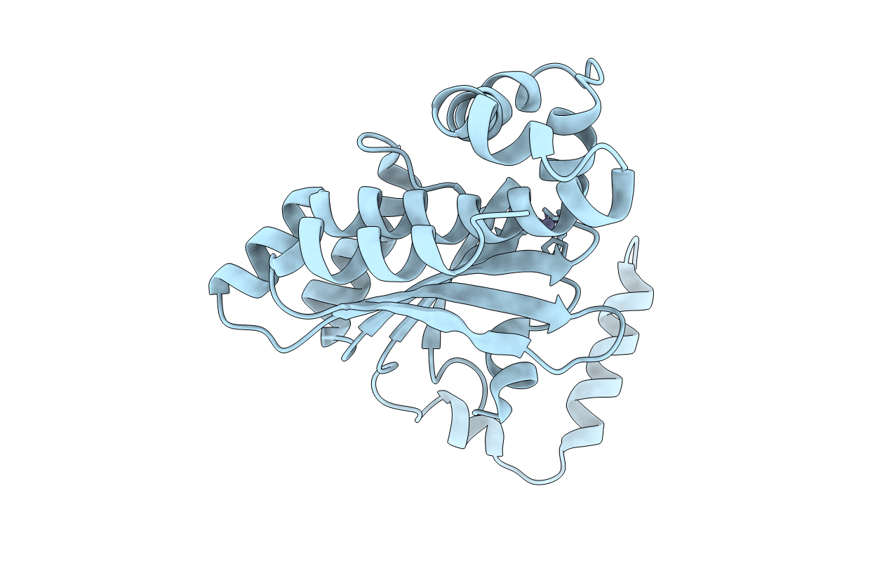
6YL7 image
Entry Detail
PDB ID:
6YL7
Keywords:
Title:
Crystal structure of beta carbonic anhydrase from the pathogenic bacterium Burkholderia pseudomallei
Biological Source:
PDB Version:
Deposition Date:
2020-04-06
Release Date:
2020-06-10
Method Details:
Experimental Method:
Resolution:
3.17 Å
R-Value Free:
0.29
R-Value Work:
0.19
R-Value Observed:
0.19
Space Group:
P 64 2 2
Macromolecular Entities
Polymer Type:polypeptide(L)
Description:Beta carbonic anhydrase
Chain IDs:A
Chain Length:256
Number of Molecules:1
Biological Source:Burkholderia pseudomallei
Ligand Molecules
Primary Citation
Crystal Structure of a Tetrameric Type II beta-Carbonic Anhydrase from the Pathogenic BacteriumBurkholderia pseudomallei.
Molecules 25 ? ? (2020)
PMID: 32408533 DOI: 10.3390/molecules25102269

Abstact

Carbonic anhydrase (CA) is a zinc enzyme that catalyzes the reversible conversion of carbon dioxide to bicarbonate and proton. Currently, CA inhibitors are widely used as antiglaucoma, anticancer, and anti-obesity drugs and for the treatment of neurological disorders. Recently, the potential use of CA inhibitors to fight infections caused by protozoa, fungi, and bacteria has emerged as a new research line. In this article, the X-ray crystal structure of β-CA from Burkholderia pseudomallei was reported. The X-ray crystal structure of this new enzyme was solved at 2.7 Å resolution, revealing a tetrameric type II β-CA with a "closed" active site in which the zinc is tetrahedrally coordinated to Cys46, Asp48, His102, and Cys105. B. pseudomallei is known to encode at least two CAs, a β-CA, and a γ-CA. These proteins, playing a pivotal role in its life cycle and pathogenicity, offer a novel therapeutic opportunity to obtain antibiotics with a different mechanism of action. Furthermore, the new structure can provide a clear view of the β-CA mechanism of action and the possibility to find selective inhibitors for this class of CAs.

Legend

Protein

Chemical

Disease

Primary Citation of related structures