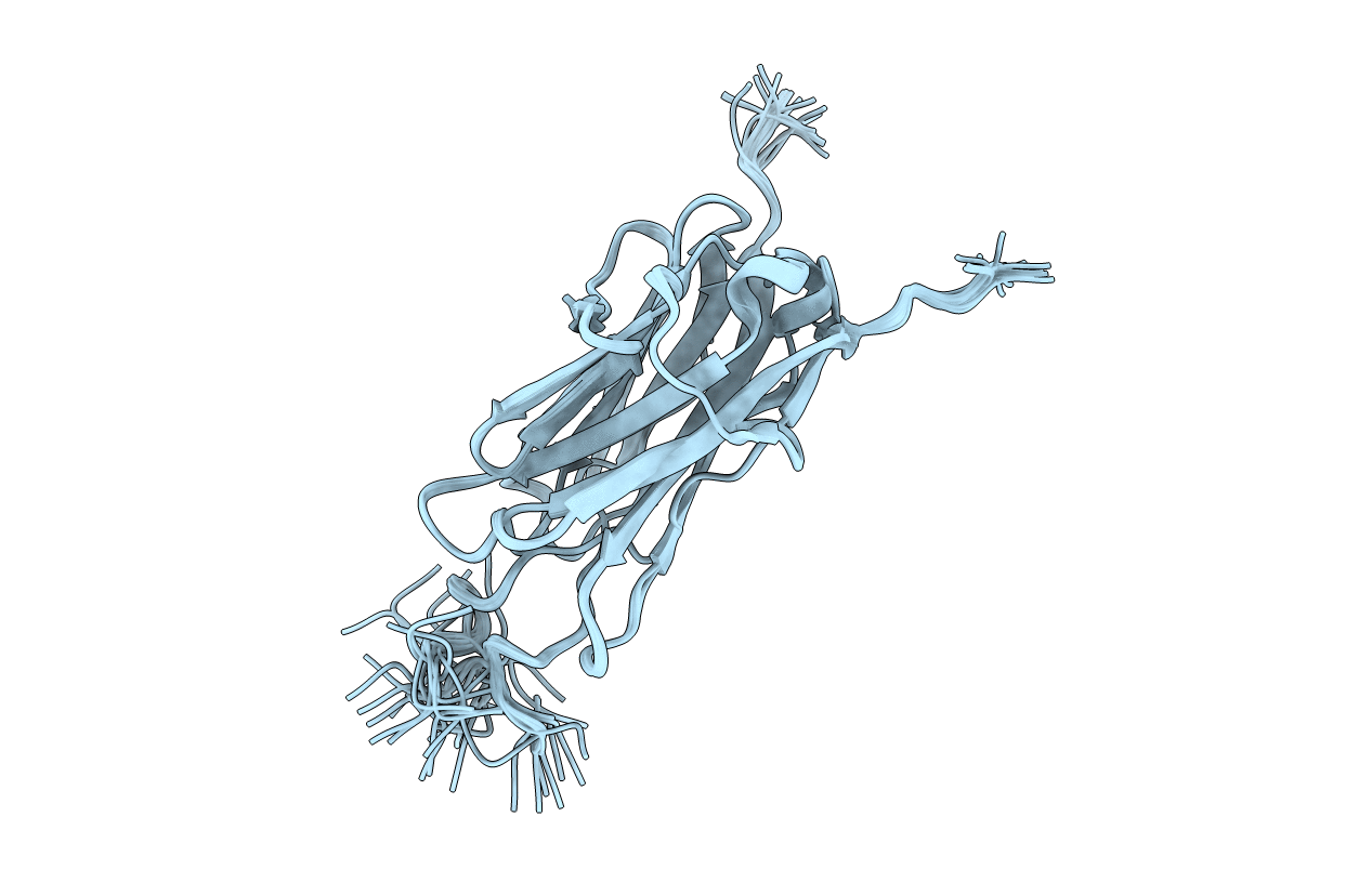
6YJ0 image
Deposition Date 2020-04-02
Release Date 2021-04-14
Last Version Date 2024-06-19
Entry Detail
PDB ID:
6YJ0
Keywords:
Title:
Solution NMR structure of titin N2A region Ig domain I83
Biological Source:
Source Organism:
Mus musculus (Taxon ID: 10090)
Host Organism:
Method Details:
Experimental Method:
Conformers Calculated:
400
Conformers Submitted:
40
Selection Criteria:
structures with the lowest energy
Macromolecular Entities
Polymer Type:polypeptide(L)
Molecule:Titin
Gene (Uniprot):Ttn
Mutations:N.A.
Chain IDs:A
Chain Length:105
Number of Molecules:1
Biological Source:Mus musculus
Ligand Molecules
Primary Citation
Solution NMR Structure of Titin N2A Region Ig Domain I83 and Its Interaction with Metal Ions.
J.Mol.Biol. 433 166977 166977 (2021)
PMID: 33811919 DOI: 10.1016/j.jmb.2021.166977

Abstact

Titin, the largest single chain protein known so far, has long been known to play a critical role in passive muscle function but recent studies have highlighted titin's role in active muscle function. One of the key elements in this role is the Ca2+-dependent interaction between titin's N2A region and the thin filament. An important element in this interaction is I83, the terminal immunoglobulin domain in the N2A region. There is limited structural information about this domain, but experimental evidence suggests that it plays a critical role in the N2A-actin binding interaction. We now report the solution NMR structure of I83 and characterize its dynamics and metal binding properties in detail. Its structure shows interesting relationships to other I-band Ig domains. Metal binding and dynamics data point towards the way the domain is evolutionarily optimized to interact with neighbouring domains. We also identify a calcium binding site on the N-terminal side of I83, which is expected to impact the interdomain interaction with the I82 domain. Together these results provide a first step towards a better understanding of the physiological effects associated with deletion of most of the I83 domain, as occurs in the mdm mouse model, as well as for future investigations of the N2A region.

Legend

Protein

Chemical

Disease

Primary Citation of related structures