6YCA image
Deposition Date 2020-03-18
Release Date 2020-09-09
Last Version Date 2024-01-31
Entry Detail
PDB ID:
6YCA
Keywords:
Title:
Crystal structure of Eis1 from Mycobacterium abscessus
Biological Source:
Source Organism:
Host Organism:
Method Details:
Experimental Method:
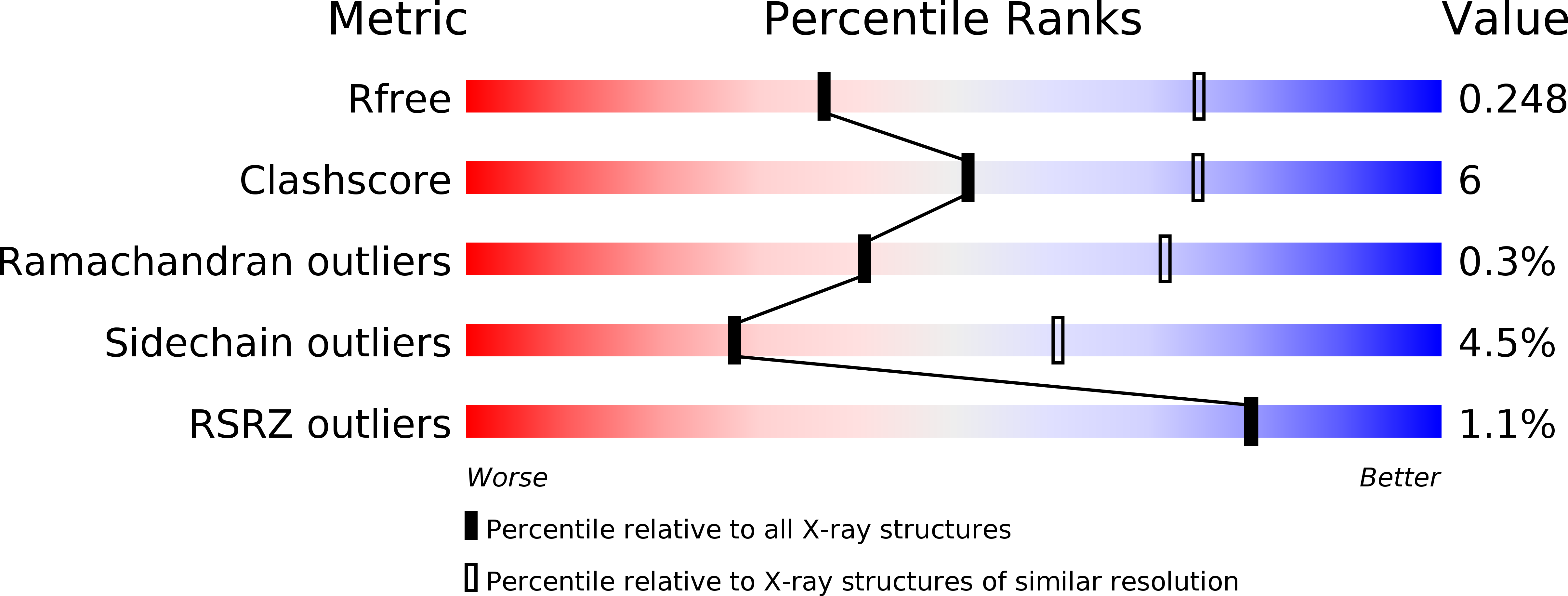
Resolution:
2.90 Å
R-Value Free:
0.24
R-Value Work:
0.20
R-Value Observed:
0.20
Space Group:
P 21 21 21
Macromolecular Entities
Polymer Type:polypeptide(L)
Molecule:Uncharacterized N-acetyltransferase D2E76_00625
Chain IDs:A, B, C, D, E, F
Chain Length:415
Number of Molecules:6
Biological Source:Mycobacteroides abscessus
Primary Citation
Structural analysis of the N-acetyltransferase Eis1 from Mycobacterium abscessus reveals the molecular determinants of its incapacity to modify aminoglycosides.
Proteins 89 94 106 (2021)
PMID: 32860271 DOI: 10.1002/prot.25997

Abstact

Enhanced intracellular survival (Eis) proteins belonging to the superfamily of the GCN5-related N-acetyltransferases play important functions in mycobacterial pathogenesis. In Mycobacterium tuberculosis, Eis enhances the intracellular survival of the bacilli in macrophages by modulating the host immune response and is capable to chemically modify and inactivate aminoglycosides. In nontuberculous mycobacteria (NTM), Eis shares similar functions. However, Mycobacterium abscessus, a multidrug resistant NTM, possesses two functionally distinct Eis homologues, Eis1Mab and Eis2Mab . While Eis2Mab participates in virulence and aminoglycosides resistance, this is not the case for Eis1Mab, whose exact biological function remains to be determined. Herein, we show that overexpression of Eis1Mab in M. abscessus fails to induce resistance to aminoglycosides. To clarify why Eis1Mab is unable to modify this class of antibiotics, we solved its crystal structure bound to its cofactor, acetyl-CoA. The structure revealed that Eis1Mab has a typical homohexameric Eis-like organization. The structural analysis supported by biochemical approaches demonstrated that while Eis1Mab can acetylate small substrates, its active site is too narrow to accommodate aminoglycosides. Comparison with other Eis structures showed that an extended loop between strands 9 and 10 is blocking the access of large substrates to the active site and movement of helices 4 and 5 reduces the volume of the substrate-binding pocket to these compounds in Eis1Mab . Overall, this study underscores the molecular determinants explaining functional differences between Eis1Mab and Eis2Mab, especially those inherent to their capacity to modify aminoglycosides.

Legend

Protein

Chemical

Disease

Primary Citation of related structures
Feedback Form
Name
Email
Institute
Feedback