
Deposition Date
2020-03-06
Release Date
2020-12-16
Last Version Date
2024-10-09
Entry Detail
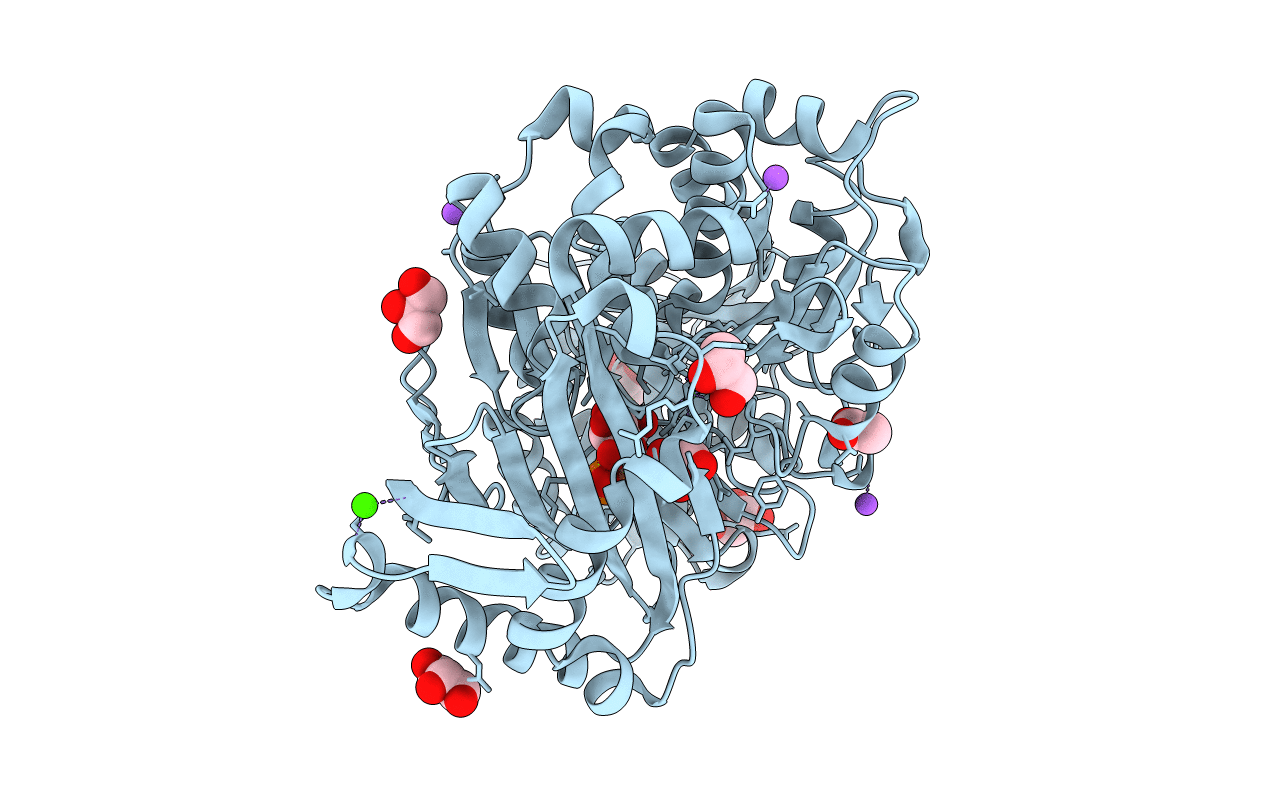
PDB ID:
6Y8Y
Keywords:
Title:
Structure of Baltic Herring (Clupea Harengus) Phosphoglucomutase 5 (PGM5) with bound Glucose-1-Phosphate
Biological Source:
Source Organism(s):
Clupea harengus (Taxon ID: 7950)
Expression System(s):
Method Details:
Experimental Method:
Resolution:
1.95 Å
R-Value Free:
0.20
R-Value Work:
0.16
R-Value Observed:
0.17
Space Group:
P 2 21 21
