
Deposition Date
2020-02-21
Release Date
2020-04-29
Last Version Date
2024-01-24
Entry Detail
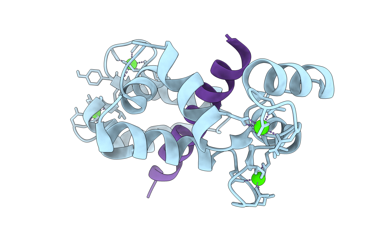
PDB ID:
6Y4O
Keywords:
Title:
Calmodulin bound to cardiac ryanodine receptor (RyR2) calmodulin binding domain
Biological Source:
Source Organism(s):
Homo sapiens (Taxon ID: 9606)
Mus musculus (Taxon ID: 10090)
Mus musculus (Taxon ID: 10090)
Expression System(s):
Method Details:
Experimental Method:
Resolution:
1.84 Å
R-Value Free:
0.20
R-Value Work:
0.17
R-Value Observed:
0.17
Space Group:
P 21 21 21
