6Y4M image
Deposition Date 2020-02-21
Release Date 2021-03-31
Last Version Date 2024-06-19
Entry Detail
PDB ID:
6Y4M
Keywords:
Title:
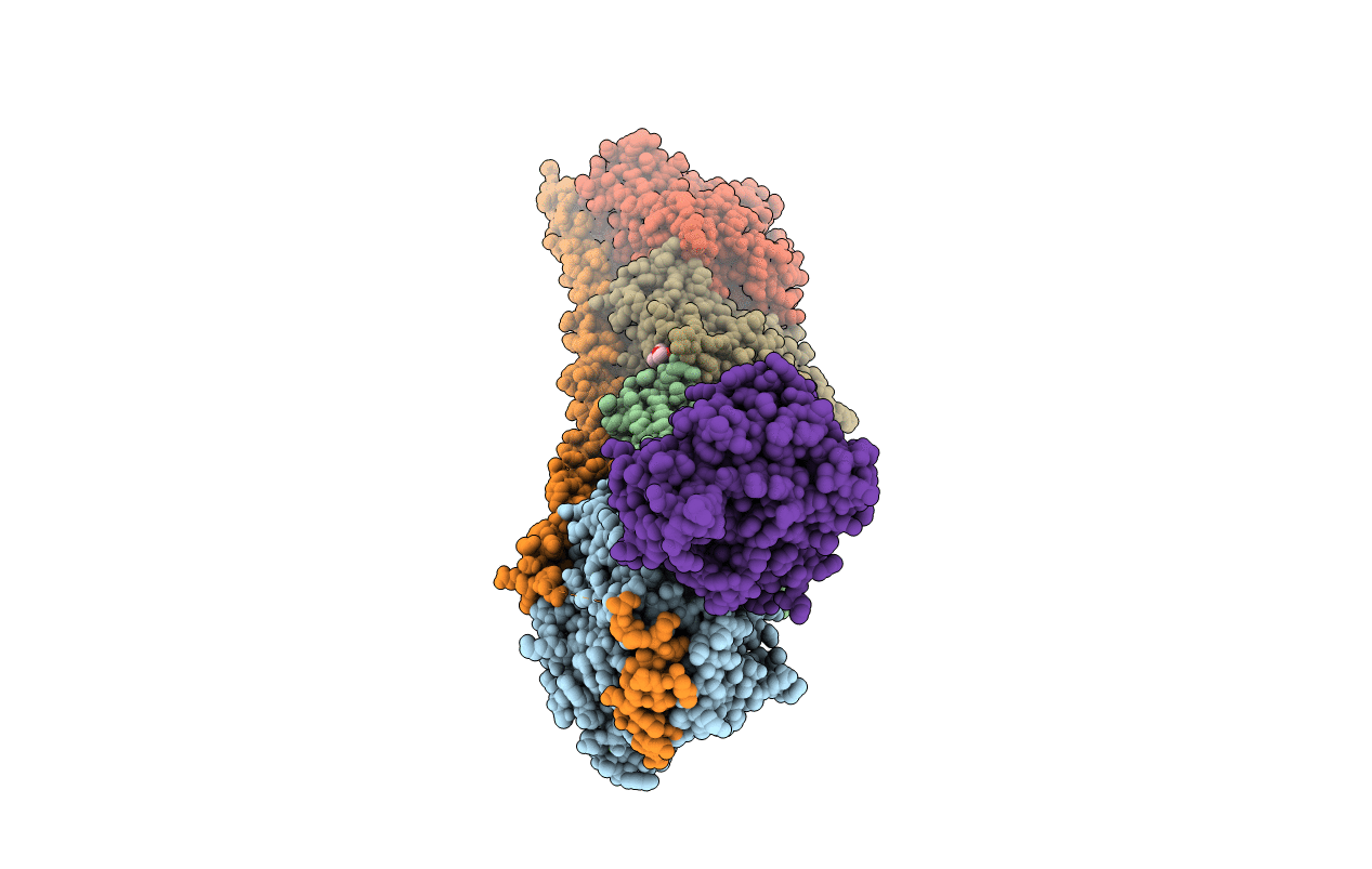
Structure of Tubulin Tyrosine Ligase in Complex with Tb111
Biological Source:
Source Organism:
Rattus norvegicus (Taxon ID: 10116)
Gallus gallus (Taxon ID: 9031)
Sus scrofa (Taxon ID: 9823)
Host Organism:
Method Details:
Experimental Method:
Resolution:
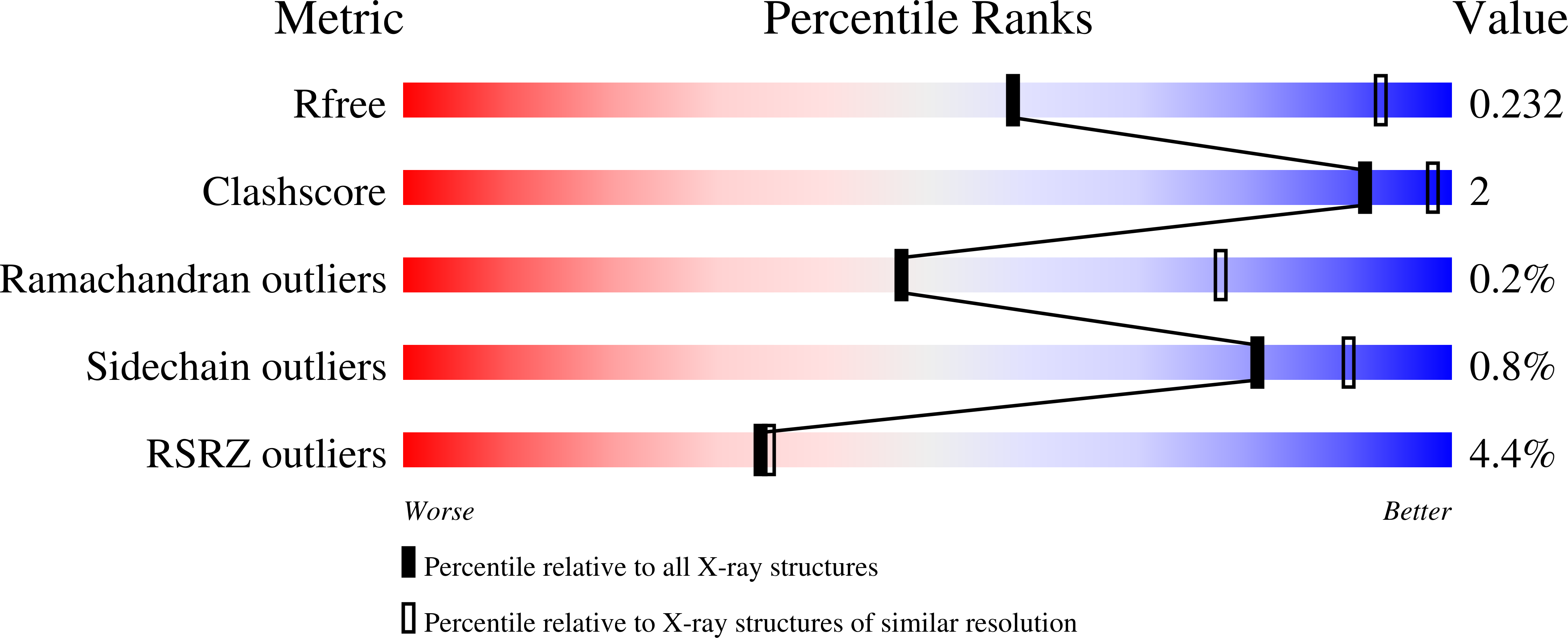
3.34 Å
R-Value Free:
0.23
R-Value Work:
0.20
R-Value Observed:
0.20
Space Group:
P 21 21 21
Macromolecular Entities
Polymer Type:polypeptide(L)
Molecule:Tubulin alpha-1B chain
Gene (Uniprot):TUBA1B
Chain IDs:A, C
Chain Length:451
Number of Molecules:2
Biological Source:Sus scrofa
Polymer Type:polypeptide(L)
Molecule:Tubulin beta chain
Chain IDs:B, D
Chain Length:445
Number of Molecules:2
Biological Source:Sus scrofa
Polymer Type:polypeptide(L)
Molecule:Stathmin-4
Gene (Uniprot):Stmn4
Chain IDs:E
Chain Length:143
Number of Molecules:1
Biological Source:Rattus norvegicus
Polymer Type:polypeptide(L)
Molecule:Tubulin-Tyrosine Ligase
Chain IDs:F
Chain Length:384
Number of Molecules:1
Biological Source:Gallus gallus
Primary Citation
Design, Synthesis, and Biological Evaluation of Tubulysin Analogues, Linker-Drugs, and Antibody-Drug Conjugates, Insights into Structure-Activity Relationships, and Tubulysin-Tubulin Binding Derived from X-ray Crystallographic Analysis.
J.Org.Chem. 86 3377 3421 (2021)
PMID: 33544599 DOI: 10.1021/acs.joc.0c02755

Abstact

Molecular design, synthesis, and biological evaluation of tubulysin analogues, linker-drugs, and antibody-drug conjugates are described. Among the new discoveries reported is the identification of new potent analogues within the tubulysin family that carry a C11 alkyl ether substituent, rather than the usual ester structural motif at that position, a fact that endows the former with higher plasma stability than that of the latter. Also described herein are X-ray crystallographic analysis studies of two tubulin-tubulysin complexes formed within the α/β interface between two tubulin heterodimers and two highly potent tubulysin analogues, one of which exhibited a different binding mode to the one previously reported for tubulysin M. The X-ray crystallographic analysis-derived new insights into the binding modes of these tubulysin analogues explain their potencies and provide inspiration for further design, synthesis, and biological investigations within this class of antitumor agents. A number of these analogues were conjugated as payloads with appropriate linkers at different sites allowing their attachment onto targeting antibodies for cancer therapies. A number of such antibody-drug conjugates were constructed and tested, both in vivo and in vitro, leading to the identification of at least one promising ADC (Herceptin-LD3), warranting further investigations.

Legend

Protein

Chemical

Disease

Primary Citation of related structures