
Deposition Date
2020-02-10
Release Date
2021-02-24
Last Version Date
2024-10-16
Entry Detail
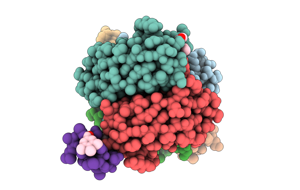
PDB ID:
6Y0V
Keywords:
Title:
Fucosylated bicyclic peptide bp71 bound to the fucose binding lectin LecB PA-IIL from Pseudomonas aeruginosa at 1.7 Angstrom resolution
Biological Source:
Source Organism(s):
Pseudomonas aeruginosa (Taxon ID: 287)
synthetic construct (Taxon ID: 32630)
synthetic construct (Taxon ID: 32630)
Expression System(s):
Method Details:
Experimental Method:
Resolution:
1.98 Å
R-Value Free:
0.23
R-Value Work:
0.17
R-Value Observed:
0.18
Space Group:
P 1 21 1
