Deposition Date
2020-02-02
Release Date
2020-03-25
Last Version Date
2024-11-20
Entry Detail
PDB ID:
6XZ6
Keywords:
Title:
Structure of the trypanosome brucei factor H receptor bound to domain D5 of bovine factor H
Biological Source:
Source Organism(s):
Trypanosoma brucei brucei TREU927 (Taxon ID: 185431)
Bos taurus (Taxon ID: 9913)
Bos taurus (Taxon ID: 9913)
Expression System(s):
Method Details:
Experimental Method:
Resolution:
2.70 Å
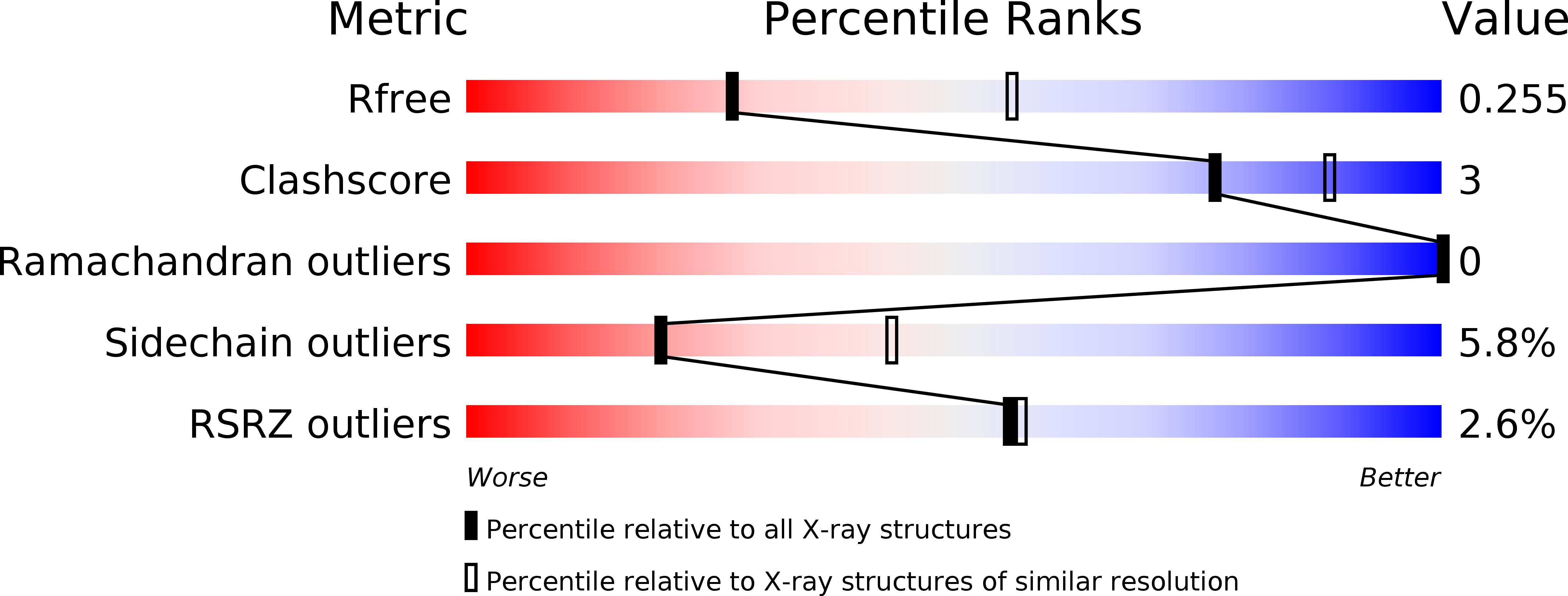
R-Value Free:
0.24
R-Value Work:
0.19
R-Value Observed:
0.19
Space Group:
C 1 2 1