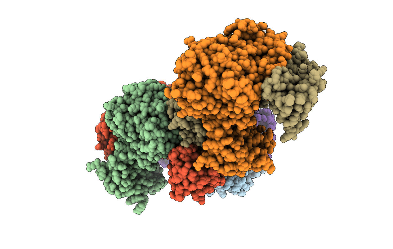
6XYL image
Entry Detail
PDB ID:
6XYL
Title:
Crystal structure of delta466-491 cystathionine beta-synthase from Toxoplasma gondii with L-serine
Biological Source:
Source Organism:
Host Organism:
PDB Version:
Deposition Date:
2020-01-30
Release Date:
2021-02-10
Method Details:
Experimental Method:
Resolution:
3.15 Å
R-Value Free:
0.29
R-Value Work:
0.27
R-Value Observed:
0.27
Space Group:
P 31
Macromolecular Entities
Polymer Type:polypeptide(L)
Description:Cystathionine beta-synthase
Chain IDs:A, B, C, D, E, F
Chain Length:514
Number of Molecules:6
Biological Source:Toxoplasma gondii ME49
Ligand Molecules
Primary Citation
Structural insight into the unique conformation of cystathionine beta-synthase from Toxoplasma gondii .
Comput Struct Biotechnol J 19 3542 3555 (2021)
PMID: 34194677 DOI: 10.1016/j.csbj.2021.05.052

Abstact

Cysteine plays a major role in the redox homeostasis and antioxidative defense mechanisms of many parasites of the phylum Apicomplexa. Of relevance to human health is Toxoplasma gondii, the causative agent of toxoplasmosis. A major route of cysteine biosynthesis in this parasite is the reverse transsulfuration pathway involving two key enzymes cystathionine β-synthase (CBS) and cystathionine γ-lyase (CGL). CBS from T. gondii (TgCBS) catalyzes the pyridoxal-5́-phosphate-dependent condensation of homocysteine with either serine or O-acetylserine to produce cystathionine. The enzyme can perform alternative reactions that use homocysteine and cysteine as substrates leading to the endogenous biosynthesis of hydrogen sulfide, another key element in maintaining the intracellular redox equilibrium. In contrast with human CBS, TgCBS lacks the N-terminal heme binding domain and is not responsive to S-adenosylmethionine. Herein, we describe the structure of a TgCBS construct that lacks amino acid residues 466-491 and shows the same activity of the native protein. TgCBS Δ466-491 was determined alone and in complex with reaction intermediates. A complementary molecular dynamics analysis revealed a unique domain organization, similar to the pathogenic mutant D444N of human CBS. Our data provides one missing piece in the structural diversity of CBSs by revealing the so far unknown three-dimensional arrangement of the CBS-type of Apicomplexa. This domain distribution is also detected in yeast and bacteria like Pseudomonas aeruginosa. These results pave the way for understanding the mechanisms by which TgCBS regulates the intracellular redox of the parasite, and have far-reaching consequences for the functional understanding of CBSs with similar domain distribution.

Legend

Protein

Chemical

Disease

Primary Citation of related structures