6XWV image
Deposition Date 2020-01-24
Release Date 2020-04-01
Last Version Date 2024-05-01
Entry Detail
PDB ID:
6XWV
Keywords:
Title:
Crystal structure of drosophila melanogaster CENP-C bound to CAL1
Biological Source:
Source Organism:
Host Organism:
Method Details:
Experimental Method:
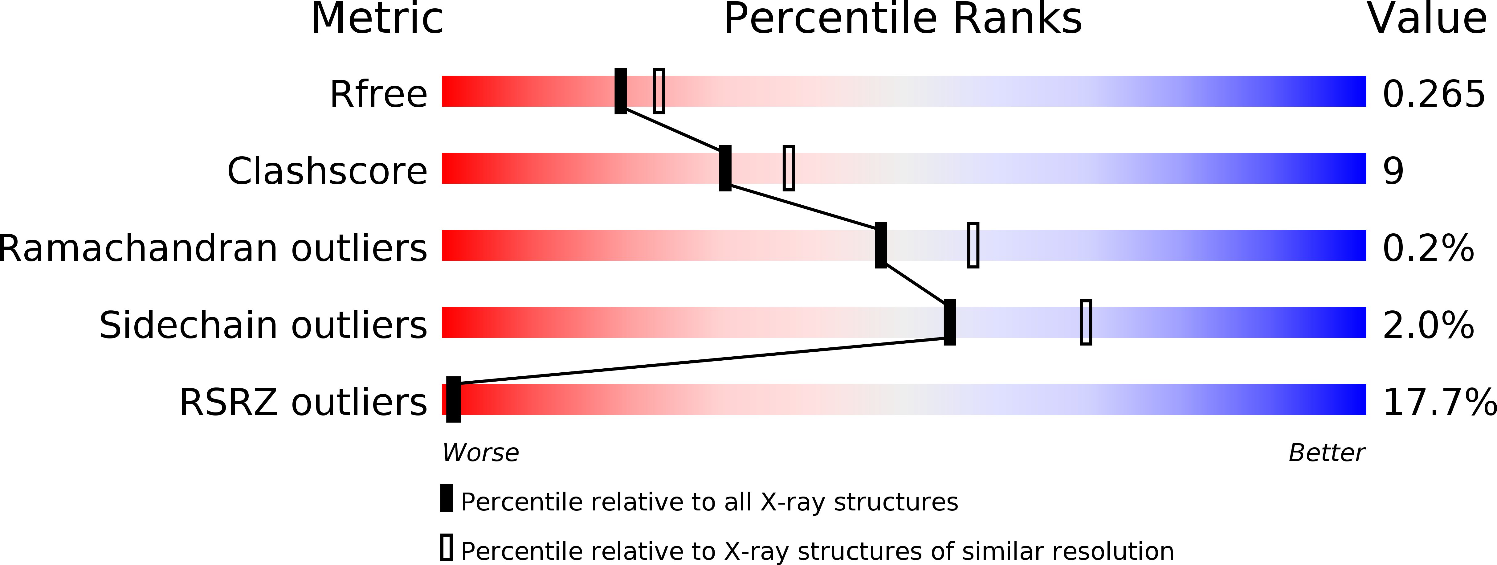
Resolution:
2.27 Å
R-Value Free:
0.26
R-Value Work:
0.23
R-Value Observed:
0.23
Space Group:
P 21 21 21
Macromolecular Entities
Structures with similar UniProt ID
Protein Blast
Polymer Type:polypeptide(L)
Molecule:Calmodulin
Chain IDs:A, C (auth: B), D (auth: C), E (auth: D)
Chain Length:1411
Number of Molecules:4
Biological Source:Drosophila melanogaster
Structures with similar UniProt ID
Protein Blast
Polymer Type:polypeptide(L)
Molecule:Ryanodine Receptor 2
Gene (Uniprot):cal1
Chain IDs:B (auth: E)
Chain Length:979
Number of Molecules:1
Biological Source:Drosophila melanogaster
Primary Citation
Structural basis for centromere maintenance by Drosophila CENP-A chaperone CAL1.
Embo J. 39 e103234 e103234 (2020)
PMID: 32134144 DOI: 10.15252/embj.2019103234

Abstact

Centromeres are microtubule attachment sites on chromosomes defined by the enrichment of histone variant CENP-A-containing nucleosomes. To preserve centromere identity, CENP-A must be escorted to centromeres by a CENP-A-specific chaperone for deposition. Despite this essential requirement, many eukaryotes differ in the composition of players involved in centromere maintenance, highlighting the plasticity of this process. In humans, CENP-A recognition and centromere targeting are achieved by HJURP and the Mis18 complex, respectively. Using X-ray crystallography, we here show how Drosophila CAL1, an evolutionarily distinct CENP-A histone chaperone, binds both CENP-A and the centromere receptor CENP-C without the requirement for the Mis18 complex. While an N-terminal CAL1 fragment wraps around CENP-A/H4 through multiple physical contacts, a C-terminal CAL1 fragment directly binds a CENP-C cupin domain dimer. Although divergent at the primary structure level, CAL1 thus binds CENP-A/H4 using evolutionarily conserved and adaptive structural principles. The CAL1 binding site on CENP-C is strategically positioned near the cupin dimerisation interface, restricting binding to just one CAL1 molecule per CENP-C dimer. Overall, by demonstrating how CAL1 binds CENP-A/H4 and CENP-C, we provide key insights into the minimalistic principles underlying centromere maintenance.

Legend

Protein

Chemical

Disease

Primary Citation of related structures