6XWH image
Deposition Date 2020-01-23
Release Date 2020-09-09
Last Version Date 2024-01-24
Entry Detail
PDB ID:
6XWH
Keywords:
Title:
Crystal Structure of the Human RXR DNA-Binding Domain Homodimer Bound to the Human Hoxb13 DR0 Response Element
Biological Source:
Source Organism:
Homo sapiens (Taxon ID: 9606)
Method Details:
Experimental Method:
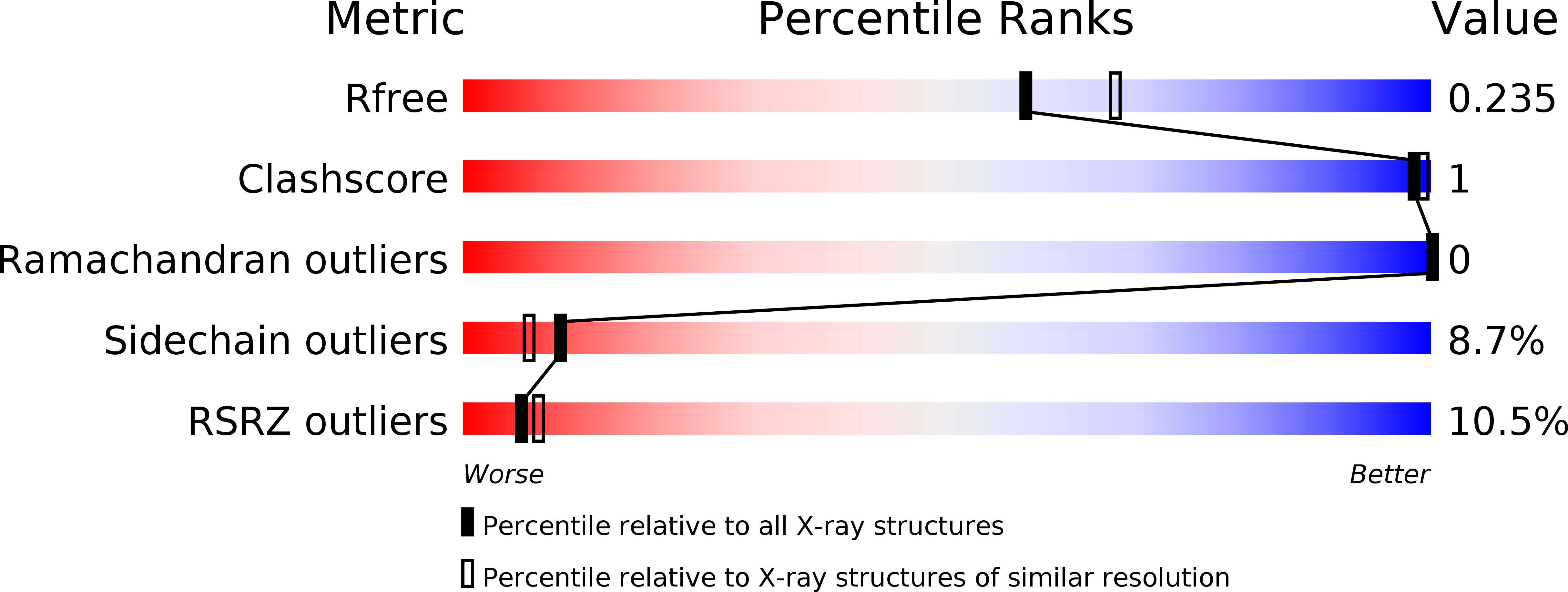
Resolution:
2.10 Å
R-Value Free:
0.22
R-Value Work:
0.18
R-Value Observed:
0.18
Space Group:
P 1 21 1
Macromolecular Entities
Polymer Type:polydeoxyribonucleotide
Molecule:Hoxb13 DR0 Response Element, 5'-3' strand
Chain IDs:A
Chain Length:16
Number of Molecules:1
Biological Source:Homo sapiens
Polymer Type:polydeoxyribonucleotide
Molecule:Hoxb13 DR0 Response Element, 3'-5' strand
Chain IDs:B
Chain Length:16
Number of Molecules:1
Biological Source:Homo sapiens
Structures with similar UniProt ID
Protein Blast
Polymer Type:polypeptide(L)
Molecule:Retinoic acid receptor RXR-alpha
Gene (Uniprot):RXRA
Chain IDs:C, D
Chain Length:87
Number of Molecules:2
Biological Source:Homo sapiens
Ligand Molecules
Primary Citation
Structural basis for DNA recognition and allosteric control of the retinoic acid receptors RAR-RXR.
Nucleic Acids Res. 48 9969 9985 (2020)
PMID: 32974652 DOI: 10.1093/nar/gkaa697

Abstact

Retinoic acid receptors (RARs) as a functional heterodimer with retinoid X receptors (RXRs), bind a diverse series of RA-response elements (RAREs) in regulated genes. Among them, the non-canonical DR0 elements are bound by RXR-RAR with comparable affinities to DR5 elements but DR0 elements do not act transcriptionally as independent RAREs. In this work, we present structural insights for the recognition of DR5 and DR0 elements by RXR-RAR heterodimer using x-ray crystallography, small angle x-ray scattering, and hydrogen/deuterium exchange coupled to mass spectrometry. We solved the crystal structures of RXR-RAR DNA-binding domain in complex with the Rarb2 DR5 and RXR-RXR DNA-binding domain in complex with Hoxb13 DR0. While cooperative binding was observed on DR5, the two molecules bound non-cooperatively on DR0 on opposite sides of the DNA. In addition, our data unveil the structural organization and dynamics of the multi-domain RXR-RAR DNA complexes providing evidence for DNA-dependent allosteric communication between domains. Differential binding modes between DR0 and DR5 were observed leading to differences in conformation and structural dynamics of the multi-domain RXR-RAR DNA complexes. These results reveal that the topological organization of the RAR binding element confer regulatory information by modulating the overall topology and structural dynamics of the RXR-RAR heterodimers.

Legend

Protein

Chemical

Disease

Primary Citation of related structures
Feedback Form
Name
Email
Institute
Feedback