6XIS image
Deposition Date 2020-06-21
Release Date 2020-10-07
Last Version Date 2024-10-16
Entry Detail
PDB ID:
6XIS
Title:
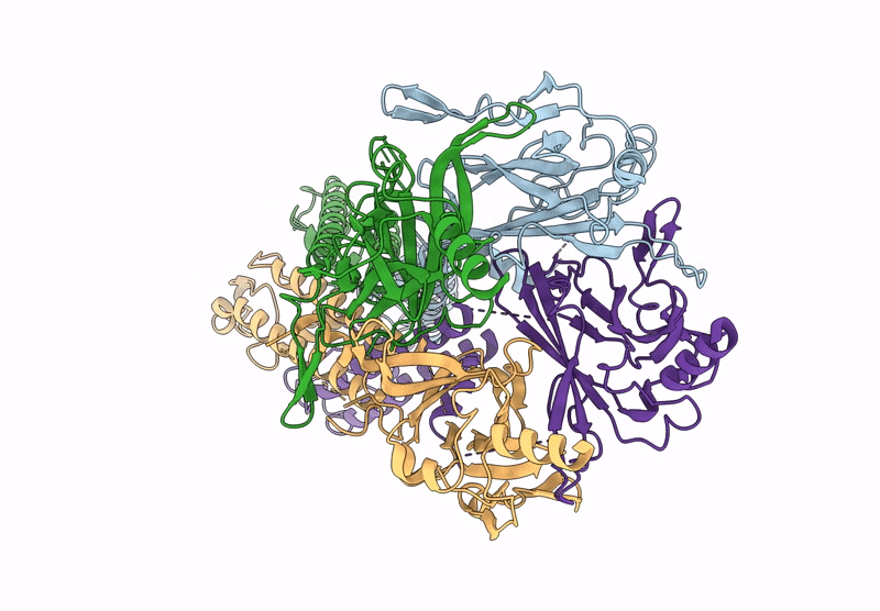
Cryo-EM structure of the G protein-gated inward rectifier K+ channel GIRK2 (Kir3.2) in apo form
Biological Source:
Source Organism:
Mus musculus (Taxon ID: 10090)
Host Organism:
Method Details:
Experimental Method:
Resolution:
3.90 Å
Aggregation State:
PARTICLE
Reconstruction Method:
SINGLE PARTICLE
Macromolecular Entities
Structures with similar UniProt ID
Protein Blast
Polymer Type:polypeptide(L)
Molecule:G protein-activated inward rectifier potassium channel 2
Chain IDs:A, B, C, D
Chain Length:340
Number of Molecules:4
Biological Source:Mus musculus
Ligand Molecules
Primary Citation
Cryo-EM analysis of PIP 2 regulation in mammalian GIRK channels.
Elife 9 ? ? (2020)
PMID: 32844743 DOI: 10.7554/eLife.60552

Abstact

G-protein-gated inward rectifier potassium (GIRK) channels are regulated by G proteins and PIP2. Here, using cryo-EM single particle analysis we describe the equilibrium ensemble of structures of neuronal GIRK2 as a function of the C8-PIP2 concentration. We find that PIP2 shifts the equilibrium between two distinguishable structures of neuronal GIRK (GIRK2), extended and docked, towards the docked form. In the docked form the cytoplasmic domain, to which Gβγ binds, becomes accessible to the cytoplasmic membrane surface where Gβγ resides. Furthermore, PIP2 binding reshapes the Gβγ binding surface on the cytoplasmic domain, preparing it to receive Gβγ. We find that cardiac GIRK (GIRK1/4) can also exist in both extended and docked conformations. These findings lead us to conclude that PIP2 influences GIRK channels in a structurally similar manner to Kir2.2 channels. In Kir2.2 channels, the PIP2-induced conformational changes open the pore. In GIRK channels, they prepare the channel for activation by Gβγ.

Legend

Protein

Chemical

Disease

Primary Citation of related structures