6XF2 image
Entry Detail
PDB ID:
6XF2
Title:
Nesprin-1G (aa2070-2200)-FHOD1(aa1-339) complex, H. sapiens
Biological Source:
Source Organism:
Host Organism:
PDB Version:
Deposition Date:
2020-06-15
Release Date:
2021-02-03
Method Details:
Experimental Method:
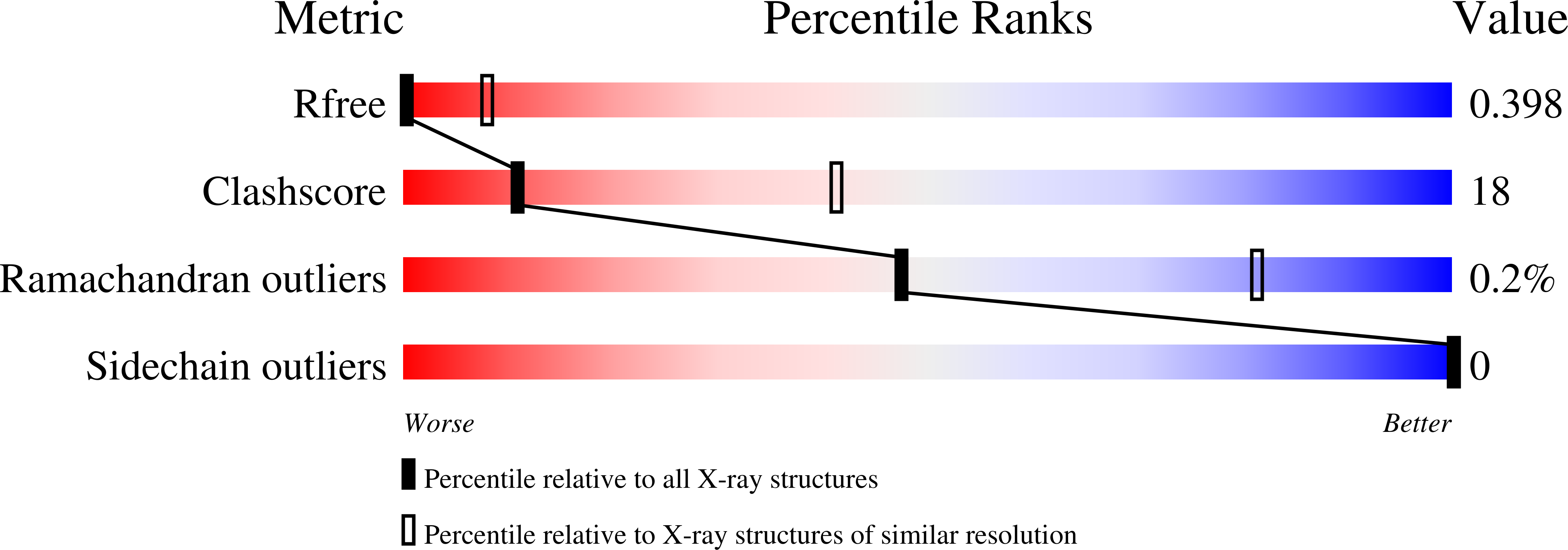
Resolution:
7.11 Å
R-Value Free:
0.39
R-Value Work:
0.33
R-Value Observed:
0.34
Space Group:
C 1 2 1
Macromolecular Entities
Polymer Type:polypeptide(L)
Description:Nesprin-1
Chain IDs:A, C
Chain Length:131
Number of Molecules:2
Biological Source:Homo sapiens
Polymer Type:polypeptide(L)
Description:FH1/FH2 domain-containing protein 1
Chain IDs:B, D
Chain Length:321
Number of Molecules:2
Biological Source:Homo sapiens
Ligand Molecules
Primary Citation
Structures of FHOD1-Nesprin1/2 complexes reveal alternate binding modes for the FH3 domain of formins.
Structure 29 540 552.e5 (2021)
PMID: 33472039 DOI: 10.1016/j.str.2020.12.013

Abstact

The nuclear position in eukaryotes is controlled by a nucleo-cytoskeletal network, critical in cell differentiation, division, and movement. Forces are transmitted through conserved Linker of Nucleoskeleton and Cytoskeleton (LINC) complexes that traverse the nuclear envelope and engage on either side of the membrane with diverse binding partners. Nesprin-2-giant (Nes2G), a LINC element in the outer nuclear membrane, connects to the actin directly as well as through FHOD1, a formin primarily involved in actin bundling. Here, we report the crystal structure of Nes2G bound to FHOD1 and show that the presumed G-binding domain of FHOD1 is rather a spectrin repeat (SR) binding enhancer for the neighboring FH3 domain. The structure reveals that SR binding by FHOD1 is likely not regulated by the diaphanous-autoregulatory domain helix of FHOD1. Finally, we establish that Nes1G also has one FHOD1 binding SR, indicating that these abundant, giant Nesprins have overlapping functions in actin-bundle recruitment for nuclear movement.

Legend

Protein

Chemical

Disease

Primary Citation of related structures