Deposition Date
2020-05-27
Release Date
2020-07-22
Last Version Date
2024-03-06
Entry Detail
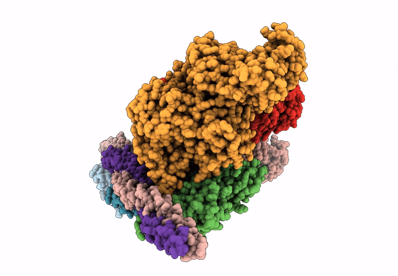
PDB ID:
6X5Z
Keywords:
Title:
Bovine Cardiac Myosin in Complex with Chicken Skeletal Actin and Human Cardiac Tropomyosin in the Rigor State
Biological Source:
Source Organism(s):
Homo sapiens (Taxon ID: 9606)
Gallus gallus (Taxon ID: 9031)
Bos taurus (Taxon ID: 9913)
Gallus gallus (Taxon ID: 9031)
Bos taurus (Taxon ID: 9913)
Expression System(s):
Method Details:
Experimental Method:
Resolution:
4.24 Å
Aggregation State:
HELICAL ARRAY
Reconstruction Method:
HELICAL