6X58 image
Deposition Date 2020-05-25
Release Date 2021-05-19
Last Version Date 2024-11-20
Entry Detail
PDB ID:
6X58
Title:
MPER-Fluc-Ec2 bound to 10E8v4 antibody
Biological Source:
Source Organism:
Host Organism:
Method Details:
Experimental Method:
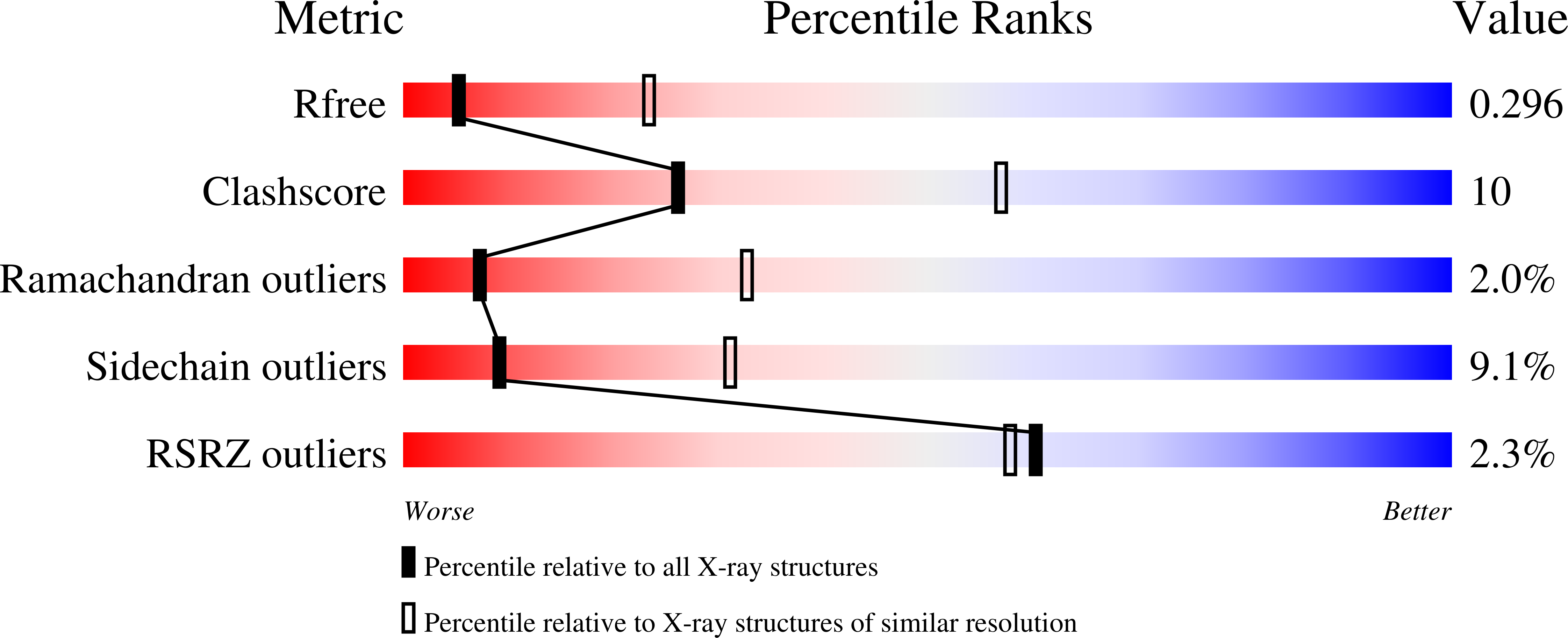
Resolution:
3.26 Å
R-Value Free:
0.29
R-Value Work:
0.25
R-Value Observed:
0.25
Space Group:
P 32
Macromolecular Entities
Polymer Type:polypeptide(L)
Molecule:10E8v4 Fab Heavy Chain
Chain IDs:A (auth: C), E (auth: A)
Chain Length:233
Number of Molecules:2
Biological Source:Homo sapiens
Polymer Type:polypeptide(L)
Molecule:10E8v4 Fab Light Chain
Chain IDs:B (auth: D), F (auth: B)
Chain Length:211
Number of Molecules:2
Biological Source:Homo sapiens
Polymer Type:polypeptide(L)
Molecule:gp41 MPER peptide,Putative fluoride ion transporter CrcB
Gene (Uniprot):crcB
Mutations:R25K,A51M in transporter
Chain IDs:C (auth: E), D (auth: F)
Chain Length:138
Number of Molecules:2
Biological Source:Human immunodeficiency virus, Escherichia coli
Ligand Molecules
Primary Citation
N-terminal Transmembrane-Helix Epitope Tag for X-ray Crystallography and Electron Microscopy of Small Membrane Proteins.
J.Mol.Biol. 433 166909 166909 (2021)
PMID: 33676924 DOI: 10.1016/j.jmb.2021.166909

Abstact

Structural studies of membrane proteins, especially small membrane proteins, are associated with well-known experimental challenges. Complexation with monoclonal antibody fragments is a common strategy to augment such proteins; however, generating antibody fragments that specifically bind a target protein is not trivial. Here we identify a helical epitope, from the membrane-proximal external region (MPER) of the gp41-transmembrane subunit of the HIV envelope protein, that is recognized by several well-characterized antibodies and that can be fused as a contiguous extension of the N-terminal transmembrane helix of a broad range of membrane proteins. To analyze whether this MPER-epitope tag might aid structural studies of small membrane proteins, we determined an X-ray crystal structure of a membrane protein target that does not crystallize without the aid of crystallization chaperones, the Fluc fluoride channel, fused to the MPER epitope and in complex with antibody. We also demonstrate the utility of this approach for single particle electron microscopy with Fluc and two additional small membrane proteins that represent different membrane protein folds, AdiC and GlpF. These studies show that the MPER epitope provides a structurally defined, rigid docking site for antibody fragments that is transferable among diverse membrane proteins and can be engineered without prior structural information. Antibodies that bind to the MPER epitope serve as effective crystallization chaperones and electron microscopy fiducial markers, enabling structural studies of challenging small membrane proteins.

Legend

Protein

Chemical

Disease

Primary Citation of related structures