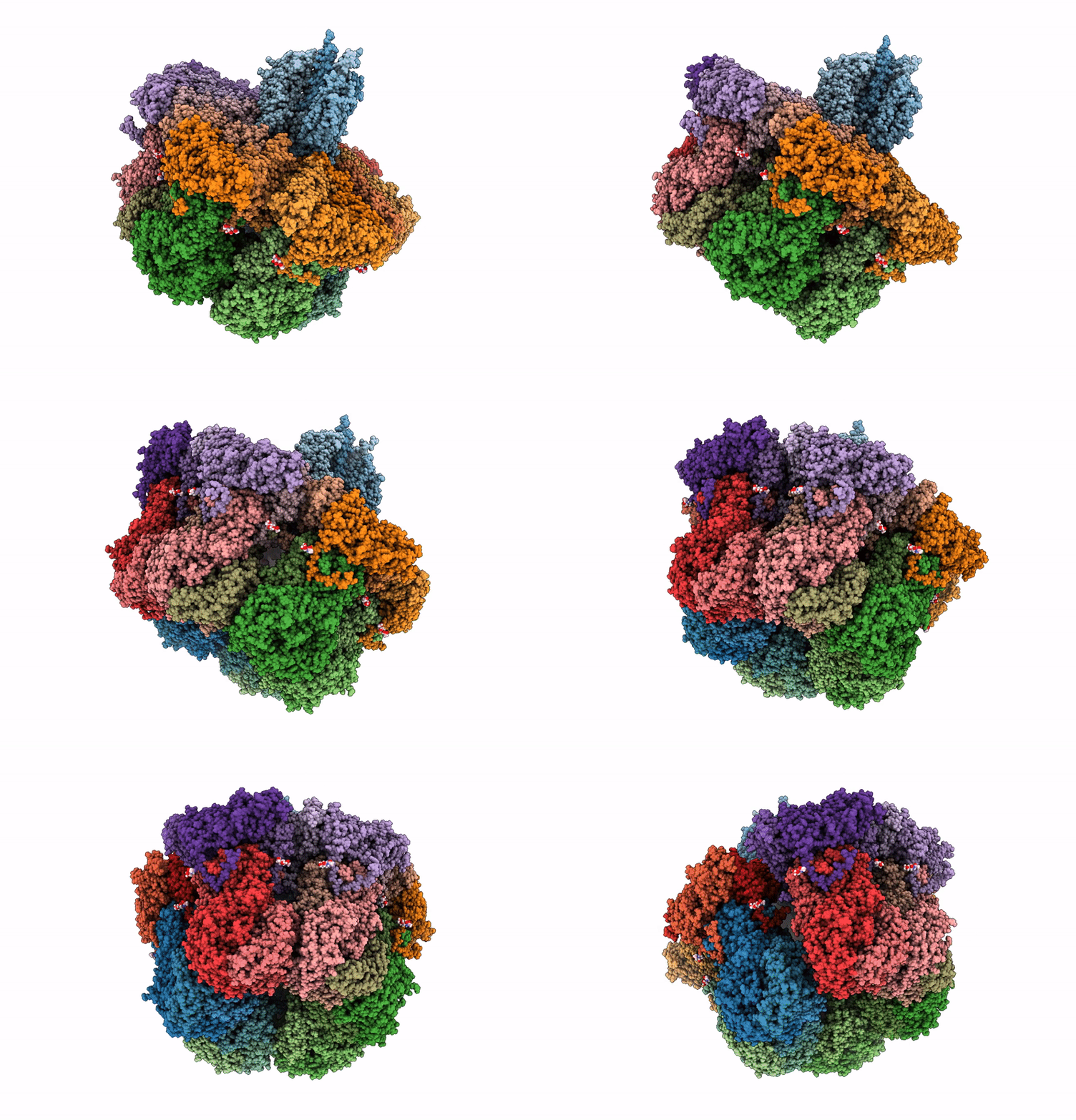
6WXG image
Entry Detail
PDB ID:
6WXG
EMDB ID:
Keywords:
Title:
Cryo-EM reconstruction of VP5*/VP8* assembly from rhesus rotavirus particles - Reversed conformation
Biological Source:
PDB Version:
Deposition Date:
2020-05-10
Release Date:
2021-01-20
Method Details:
Experimental Method:
Resolution:
3.30 Å
Aggregation State:
PARTICLE
Reconstruction Method:
SINGLE PARTICLE
Macromolecular Entities
Polymer Type:polypeptide(L)
Description:Outer capsid protein VP4
Chain IDs:A (auth: 1), B (auth: 2), C (auth: 3)
Chain Length:776
Number of Molecules:3
Biological Source:Rotavirus A (strain RVA/Monkey/United States/RRV/1975/G3P5B[3])
Polymer Type:polypeptide(L)
Description:Intermediate capsid protein VP6
Chain IDs:D (auth: A), E (auth: B), F (auth: C), G (auth: D), H (auth: E), I (auth: F), J (auth: G), K (auth: H), L (auth: I), M (auth: J), N (auth: K), O (auth: L), P (auth: M), Q (auth: N), R (auth: O), S (auth: P), T (auth: Q), U (auth: R)
Chain Length:397
Number of Molecules:18
Biological Source:Rotavirus A (strain RVA/Monkey/United States/RRV/1975/G3P5B[3])
Polymer Type:polypeptide(L)
Description:Outer capsid glycoprotein VP7
Chain IDs:V (auth: a), W (auth: b), X (auth: c), Y (auth: d), Z (auth: e), AA (auth: f), BA (auth: g), CA (auth: h), DA (auth: i), EA (auth: j), FA (auth: k), GA (auth: l), HA (auth: m), IA (auth: n), JA (auth: o), KA (auth: p), LA (auth: q), MA (auth: r)
Chain Length:397
Number of Molecules:18
Biological Source:Rotavirus A (strain RVA/Monkey/United States/RRV/1975/G3P5B[3])
Primary Citation
Functional refolding of the penetration protein on a non-enveloped virus.
Nature 590 666 670 (2021)
PMID: 33442061 DOI: 10.1038/s41586-020-03124-4

Abstact

A non-enveloped virus requires a membrane lesion to deliver its genome into a target cell1. For rotaviruses, membrane perforation is a principal function of the viral outer-layer protein, VP42,3. Here we describe the use of electron cryomicroscopy to determine how VP4 performs this function and show that when activated by cleavage to VP8* and VP5*, VP4 can rearrange on the virion surface from an 'upright' to a 'reversed' conformation. The reversed structure projects a previously buried 'foot' domain outwards into the membrane of the host cell to which the virion has attached. Electron cryotomograms of virus particles entering cells are consistent with this picture. Using a disulfide mutant of VP4, we have also stabilized a probable intermediate in the transition between the two conformations. Our results define molecular mechanisms for the first steps of the penetration of rotaviruses into the membranes of target cells and suggest similarities with mechanisms postulated for other viruses.

Legend

Protein

Chemical

Disease

Primary Citation of related structures