Deposition Date
2020-05-09
Release Date
2021-05-05
Last Version Date
2024-05-29
Entry Detail
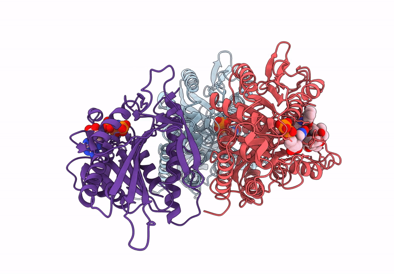
Biological Source:
Source Organism(s):
Mus musculus (Taxon ID: 10090)
Sus scrofa (Taxon ID: 9823)
Sus scrofa (Taxon ID: 9823)
Expression System(s):
Method Details:
Experimental Method:
Resolution:
3.10 Å
Aggregation State:
FILAMENT
Reconstruction Method:
HELICAL