Deposition Date
2020-05-05
Release Date
2020-11-11
Last Version Date
2024-10-16
Entry Detail
PDB ID:
6WV8
Keywords:
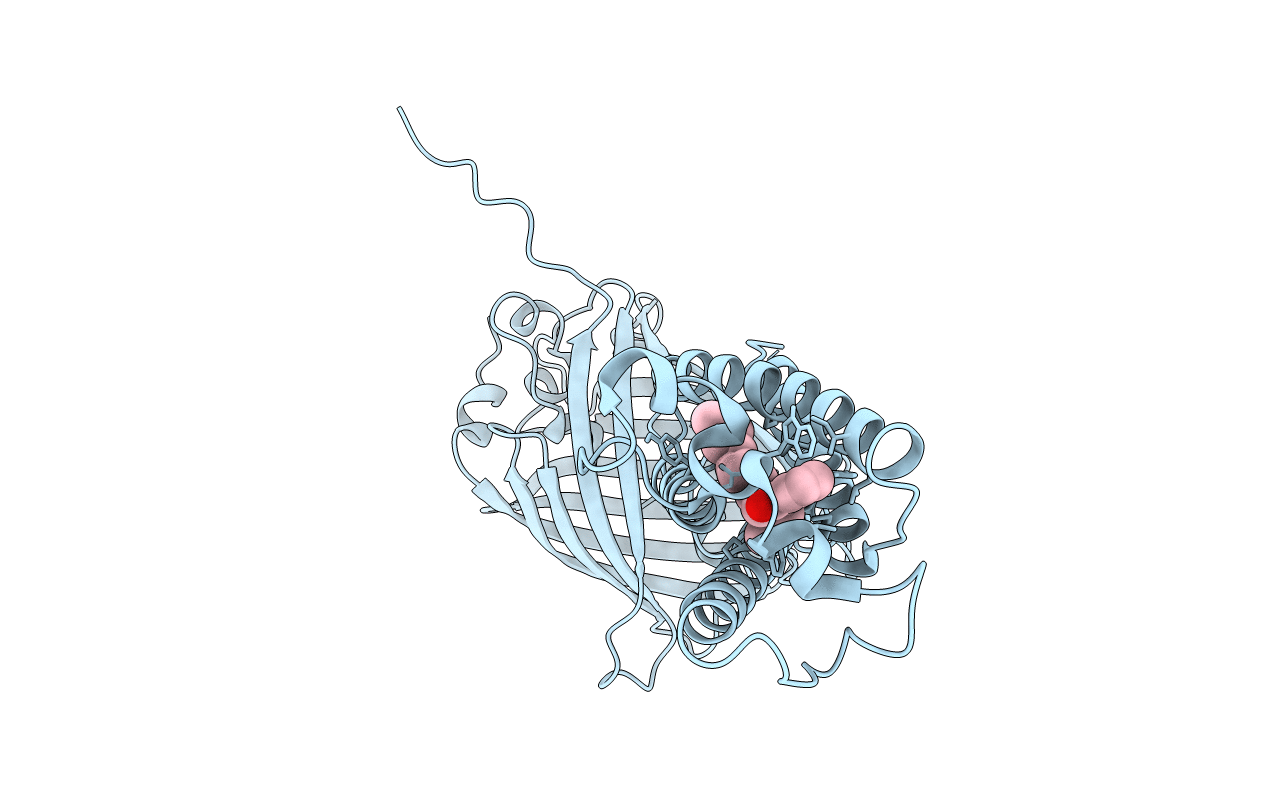
Title:
Takifugu rubripes VKOR-like C138S mutant with vitamin K1
Biological Source:
Source Organism(s):
Escherichia virus RB43 (Taxon ID: 115991)
Takifugu rubripes (Taxon ID: 31033)
Aequorea victoria (Taxon ID: 6100)
Takifugu rubripes (Taxon ID: 31033)
Aequorea victoria (Taxon ID: 6100)
Expression System(s):
Method Details:
Experimental Method:
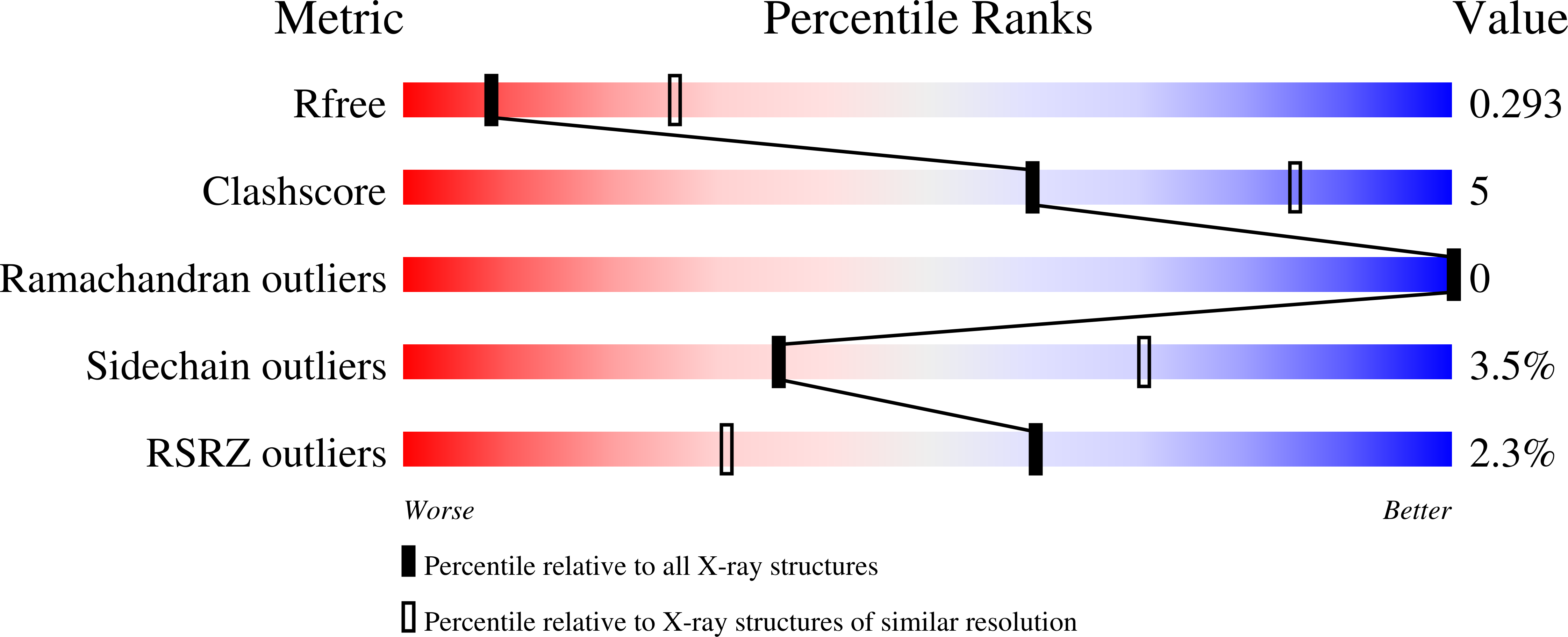
Resolution:
3.01 Å
R-Value Free:
0.29
R-Value Work:
0.25
R-Value Observed:
0.25
Space Group:
P 21 21 21