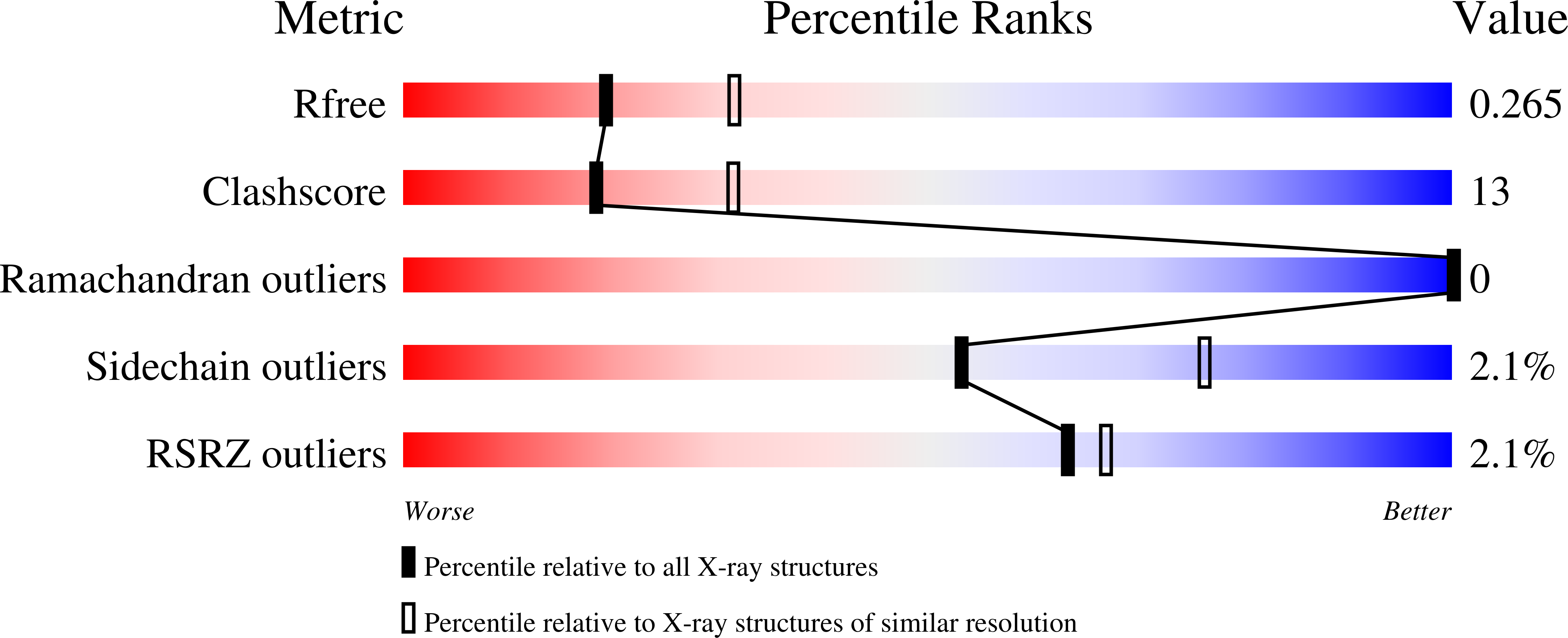
6WQX image
Deposition Date 2020-04-29
Release Date 2021-02-17
Last Version Date 2023-10-18
Entry Detail
PDB ID:
6WQX
Title:
Human PRPK-TPRKB complex
Biological Source:
Source Organism:
Homo sapiens (Taxon ID: 9606)
Host Organism:
Method Details:
Experimental Method:
Resolution:
2.53 Å
R-Value Free:
0.26
R-Value Work:
0.21
R-Value Observed:
0.21
Space Group:
P 1 21 1
Macromolecular Entities
Polymer Type:polypeptide(L)
Molecule:EKC/KEOPS complex subunit TPRKB
Gene (Uniprot):TPRKB
Chain IDs:A (auth: B), C (auth: A)
Chain Length:179
Number of Molecules:2
Biological Source:Homo sapiens
Polymer Type:polypeptide(L)
Molecule:EKC/KEOPS complex subunit TP53RK
Gene (Uniprot):TP53RK
Chain IDs:B (auth: C), D
Chain Length:267
Number of Molecules:2
Biological Source:Homo sapiens
Primary Citation
Crystal structure of the human PRPK-TPRKB complex.
Commun Biol 4 167 167 (2021)
PMID: 33547416 DOI: 10.1038/s42003-021-01683-4

Abstact

Mutations of the p53-related protein kinase (PRPK) and TP53RK-binding protein (TPRKB) cause Galloway-Mowat syndrome (GAMOS) and are found in various human cancers. We have previously shown that small compounds targeting PRPK showed anti-cancer activity against colon and skin cancer. Here we present the 2.53 Å crystal structure of the human PRPK-TPRKB-AMPPNP (adenylyl-imidodiphosphate) complex. The structure reveals details in PRPK-AMPPNP coordination and PRPK-TPRKB interaction. PRPK appears in an active conformation, albeit lacking the conventional kinase activation loop. We constructed a structural model of the human EKC/KEOPS complex, composed of PRPK, TPRKB, OSGEP, LAGE3, and GON7. Disease mutations in PRPK and TPRKB are mapped into the structure, and we show that one mutation, PRPK K238Nfs*2, lost the binding to OSGEP. Our structure also makes the virtual screening possible and paves the way for more rational drug design.

Legend

Protein

Chemical

Disease

Primary Citation of related structures