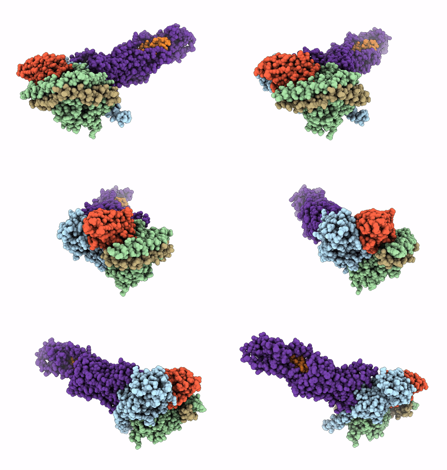
6WPW image
Deposition Date 2020-04-28
Release Date 2020-08-12
Last Version Date 2024-10-30
Entry Detail
PDB ID:
6WPW
Title:
GCGR-Gs signaling complex bound to a designed glucagon derivative
Biological Source:
Source Organism:
Homo sapiens (Taxon ID: 9606)
Lama glama (Taxon ID: 9844)
synthetic construct (Taxon ID: 32630)
Host Organism:
Method Details:
Experimental Method:
Resolution:
3.10 Å
Aggregation State:
PARTICLE
Reconstruction Method:
SINGLE PARTICLE
Macromolecular Entities
Structures with similar UniProt ID
Protein Blast
Polymer Type:polypeptide(L)
Molecule:Guanine nucleotide-binding protein G(s) subunit alpha isoforms short
Gene (Uniprot):GNAS
Chain IDs:A (auth: C)
Chain Length:380
Number of Molecules:1
Biological Source:Homo sapiens
Structures with similar UniProt ID
Protein Blast
Polymer Type:polypeptide(L)
Molecule:Guanine nucleotide-binding protein G(I)/G(S)/G(T) subunit beta-1
Gene (Uniprot):GNB1
Chain IDs:B (auth: D)
Chain Length:358
Number of Molecules:1
Biological Source:Homo sapiens
Structures with similar UniProt ID
Protein Blast
Polymer Type:polypeptide(L)
Molecule:Guanine nucleotide-binding protein G(I)/G(S)/G(O) subunit gamma-2
Gene (Uniprot):GNG2
Chain IDs:C (auth: G)
Chain Length:71
Number of Molecules:1
Biological Source:Homo sapiens
Polymer Type:polypeptide(L)
Molecule:Nb35
Chain IDs:D (auth: N)
Chain Length:138
Number of Molecules:1
Biological Source:Lama glama
Polymer Type:polypeptide(L)
Molecule:Glucagon derivative ZP3780
Chain IDs:E (auth: P)
Chain Length:29
Number of Molecules:1
Biological Source:synthetic construct
Structures with similar UniProt ID
Protein Blast
Polymer Type:polypeptide(L)
Molecule:Glucagon receptor
Gene (Uniprot):GCGR
Chain IDs:F (auth: R)
Chain Length:496
Number of Molecules:1
Biological Source:Homo sapiens
Ligand Molecules
Primary Citation
Structural insights into differences in G protein activation by family A and family B GPCRs.
Science 369 ? ? (2020)
PMID: 32732395 DOI: 10.1126/science.aba3373

Abstact

Family B heterotrimeric guanine nucleotide-binding protein (G protein)-coupled receptors (GPCRs) play important roles in carbohydrate metabolism. Recent structures of family B GPCR-Gs protein complexes reveal a disruption in the α-helix of transmembrane segment 6 (TM6) not observed in family A GPCRs. To investigate the functional impact of this structural difference, we compared the structure and function of the glucagon receptor (GCGR; family B) with the β2 adrenergic receptor (β2AR; family A). We determined the structure of the GCGR-Gs complex by means of cryo-electron microscopy at 3.1-angstrom resolution. This structure shows the distinct break in TM6. Guanosine triphosphate (GTP) turnover, guanosine diphosphate release, GTP binding, and G protein dissociation studies revealed much slower rates for G protein activation by the GCGR compared with the β2AR. Fluorescence and double electron-electron resonance studies suggest that this difference is due to the inability of agonist alone to induce a detectable outward movement of the cytoplasmic end of TM6.

Legend

Protein

Chemical

Disease

Primary Citation of related structures