6WNL image
Deposition Date 2020-04-22
Release Date 2020-07-01
Last Version Date 2024-04-03
Entry Detail
PDB ID:
6WNL
Title:
human Artemis/SNM1C catalytic domain, crystal form 2
Biological Source:
Source Organism:
Homo sapiens (Taxon ID: 9606)
Host Organism:
Method Details:
Experimental Method:
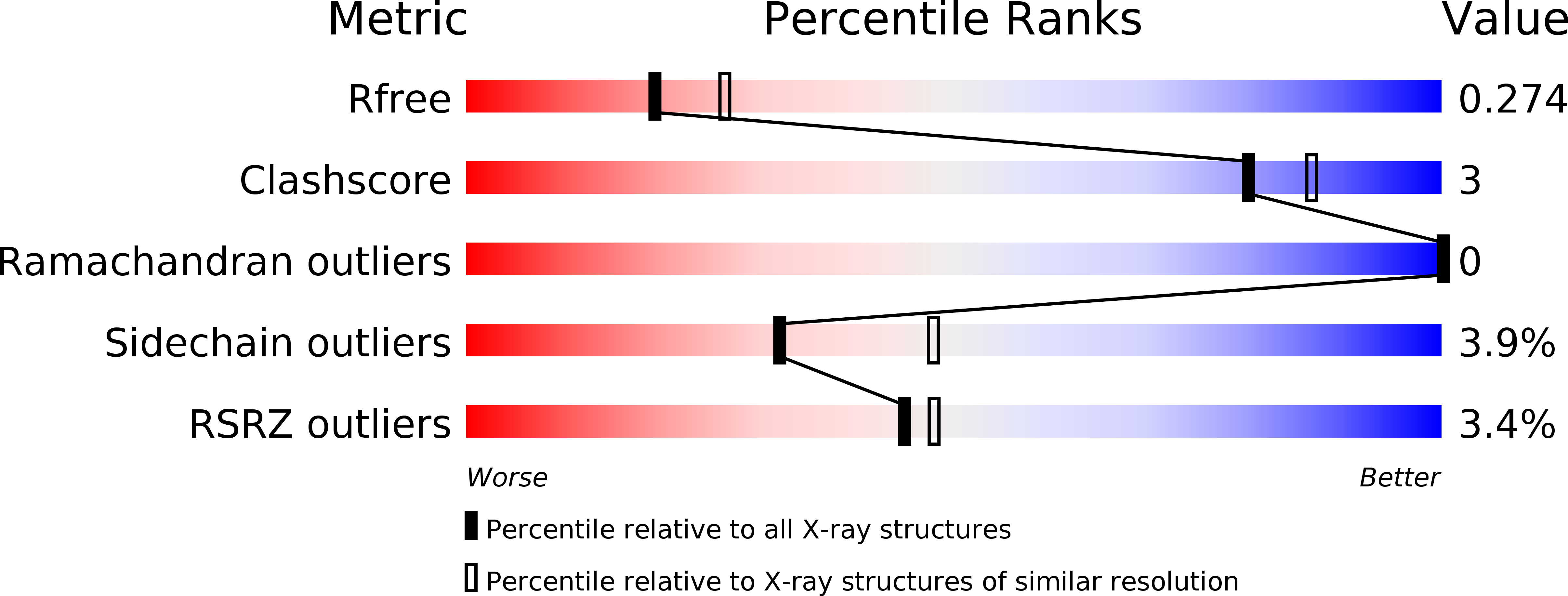
Resolution:
2.37 Å
R-Value Free:
0.28
R-Value Work:
0.22
Space Group:
P 21 21 21
Macromolecular Entities
Polymer Type:polypeptide(L)
Molecule:Protein artemis
Gene (Uniprot):DCLRE1C
Chain IDs:A, B
Chain Length:375
Number of Molecules:2
Biological Source:Homo sapiens
Ligand Molecules
Primary Citation
Structural analysis of the catalytic domain of Artemis endonuclease/SNM1C reveals distinct structural features.
J.Biol.Chem. 295 12368 12377 (2020)
PMID: 32576658 DOI: 10.1074/jbc.RA120.014136

Abstact

The endonuclease Artemis is responsible for opening DNA hairpins during V(D)J recombination and for processing a subset of pathological DNA double-strand breaks. Artemis is an attractive target for the development of therapeutics to manage various B cell and T cell tumors, because failure to open DNA hairpins and accumulation of chromosomal breaks may reduce the proliferation and viability of pre-T and pre-B cell derivatives. However, structure-based drug discovery of specific Artemis inhibitors has been hampered by a lack of crystal structures. Here, we report the structure of the catalytic domain of recombinant human Artemis. The catalytic domain displayed a polypeptide fold similar overall to those of other members in the DNA cross-link repair gene SNM1 family and in mRNA 3'-end-processing endonuclease CPSF-73, containing metallo-β-lactamase and β-CASP domains and a cluster of conserved histidine and aspartate residues capable of binding two metal atoms in the catalytic site. As in SNM1A, only one zinc ion was located in the Artemis active site. However, Artemis displayed several unique features. Unlike in other members of this enzyme class, a second zinc ion was present in the β-CASP domain that leads to structural reorientation of the putative DNA-binding surface and extends the substrate-binding pocket to a new pocket, pocket III. Moreover, the substrate-binding surface exhibited a dominant and extensive positive charge distribution compared with that in the structures of SNM1A and SNM1B, presumably because of the structurally distinct DNA substrate of Artemis. The structural features identified here may provide opportunities for designing selective Artemis inhibitors.

Legend

Protein

Chemical

Disease

Primary Citation of related structures