Deposition Date
2020-04-22
Release Date
2020-07-29
Last Version Date
2024-10-30
Entry Detail
PDB ID:
6WN4
Keywords:
Title:
Structural basis for the binding of monoclonal antibody 5D2 to the tryptophan-rich lipid-binding loop in lipoprotein lipase
Biological Source:
Source Organism(s):
Homo sapiens (Taxon ID: 9606)
Mus musculus (Taxon ID: 10090)
Mus musculus (Taxon ID: 10090)
Method Details:
Experimental Method:
Resolution:
2.80 Å
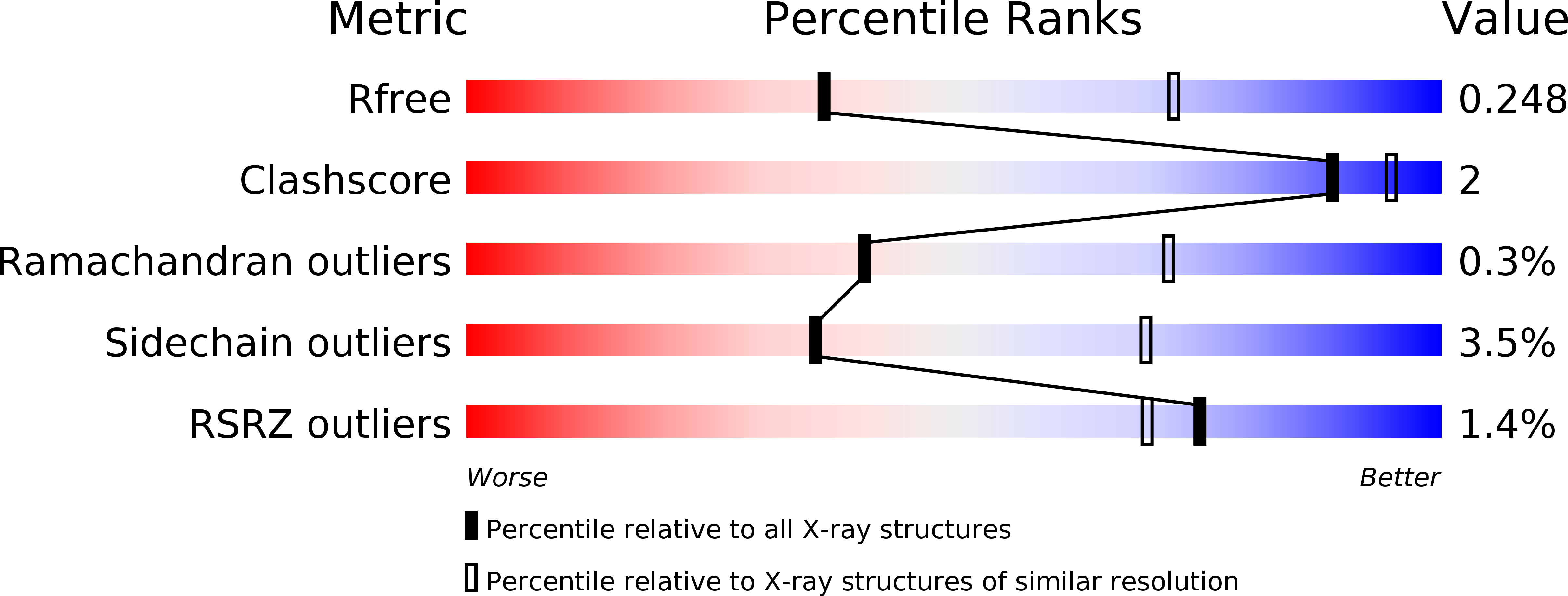
R-Value Free:
0.24
R-Value Work:
0.20
Space Group:
P 1 21 1