6WMM image
Deposition Date 2020-04-21
Release Date 2020-12-02
Last Version Date 2024-10-30
Entry Detail
PDB ID:
6WMM
Keywords:
Title:
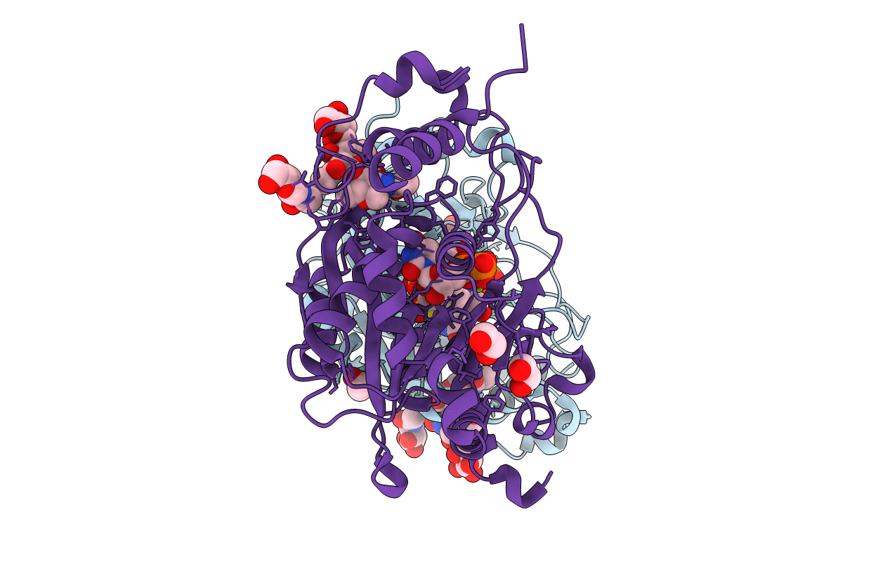
Human poly-N-acetyl-lactosamine synthase structure demonstrates a modular assembly of catalytic subsites for GT-A glycosyltransferases
Biological Source:
Source Organism:
Homo sapiens (Taxon ID: 9606)
Host Organism:
Method Details:
Experimental Method:
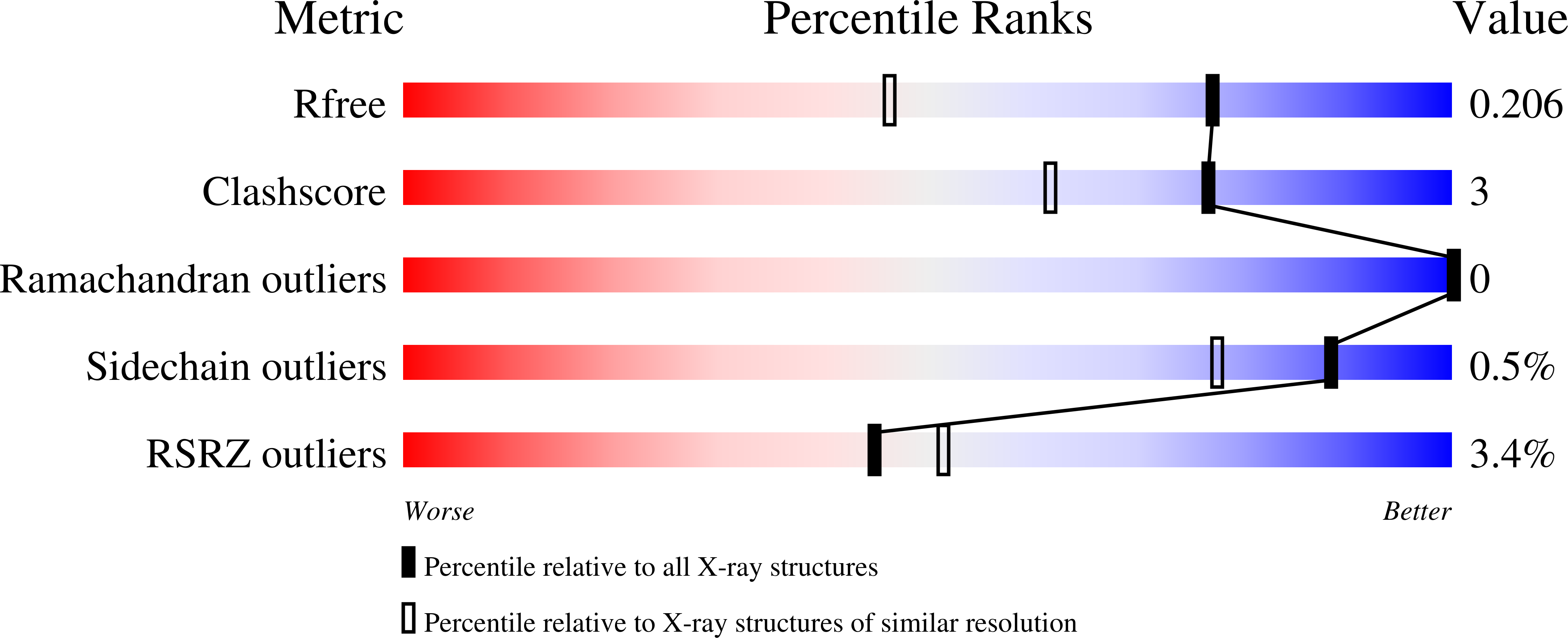
Resolution:
1.55 Å
R-Value Free:
0.19
R-Value Work:
0.17
R-Value Observed:
0.17
Space Group:
P 21 21 21
Macromolecular Entities
Polymer Type:polypeptide(L)
Molecule:N-acetyllactosaminide beta-1,3-N-acetylglucosaminyltransferase 2
Gene (Uniprot):B3GNT2
Chain IDs:A, B
Chain Length:364
Number of Molecules:2
Biological Source:Homo sapiens
Modified Residue
Compound ID Chain ID Parent Comp ID Details 2D Image
MSE A MET modified residue
Primary Citation
Comparison of human poly-N-acetyl-lactosamine synthase structure with GT-A fold glycosyltransferases supports a modular assembly of catalytic subsites.
J.Biol.Chem. 296 100110 100110 (2020)
PMID: 33229435 DOI: 10.1074/jbc.RA120.015305

Abstact

Poly-N-acetyl-lactosamine (poly-LacNAc) structures are composed of repeating [-Galβ(1,4)-GlcNAcβ(1,3)-]n glycan extensions. They are found on both N- and O-glycoproteins and glycolipids and play an important role in development, immune function, and human disease. The majority of mammalian poly-LacNAc is synthesized by the alternating iterative action of β1,3-N-acetylglucosaminyltransferase 2 (B3GNT2) and β1,4-galactosyltransferases. B3GNT2 is in the largest mammalian glycosyltransferase family, GT31, but little is known about the structure, substrate recognition, or catalysis by family members. Here we report the structures of human B3GNT2 in complex with UDP:Mg2+ and in complex with both UDP:Mg2+ and a glycan acceptor, lacto-N-neotetraose. The B3GNT2 structure conserves the GT-A fold and the DxD motif that coordinates a Mg2+ ion for binding the UDP-GlcNAc sugar donor. The acceptor complex shows interactions with only the terminal Galβ(1,4)-GlcNAcβ(1,3)- disaccharide unit, which likely explains the specificity for both N- and O-glycan acceptors. Modeling of the UDP-GlcNAc donor supports a direct displacement inverting catalytic mechanism. Comparative structural analysis indicates that nucleotide sugar donors for GT-A fold glycosyltransferases bind in similar positions and conformations without conserving interacting residues, even for enzymes that use the same donor substrate. In contrast, the B3GNT2 acceptor binding site is consistent with prior models suggesting that the evolution of acceptor specificity involves loops inserted into the stable GT-A fold. These observations support the hypothesis that GT-A fold glycosyltransferases employ coevolving donor, acceptor, and catalytic subsite modules as templates to achieve the complex diversity of glycan linkages in biological systems.

Legend

Protein

Chemical

Disease

Primary Citation of related structures