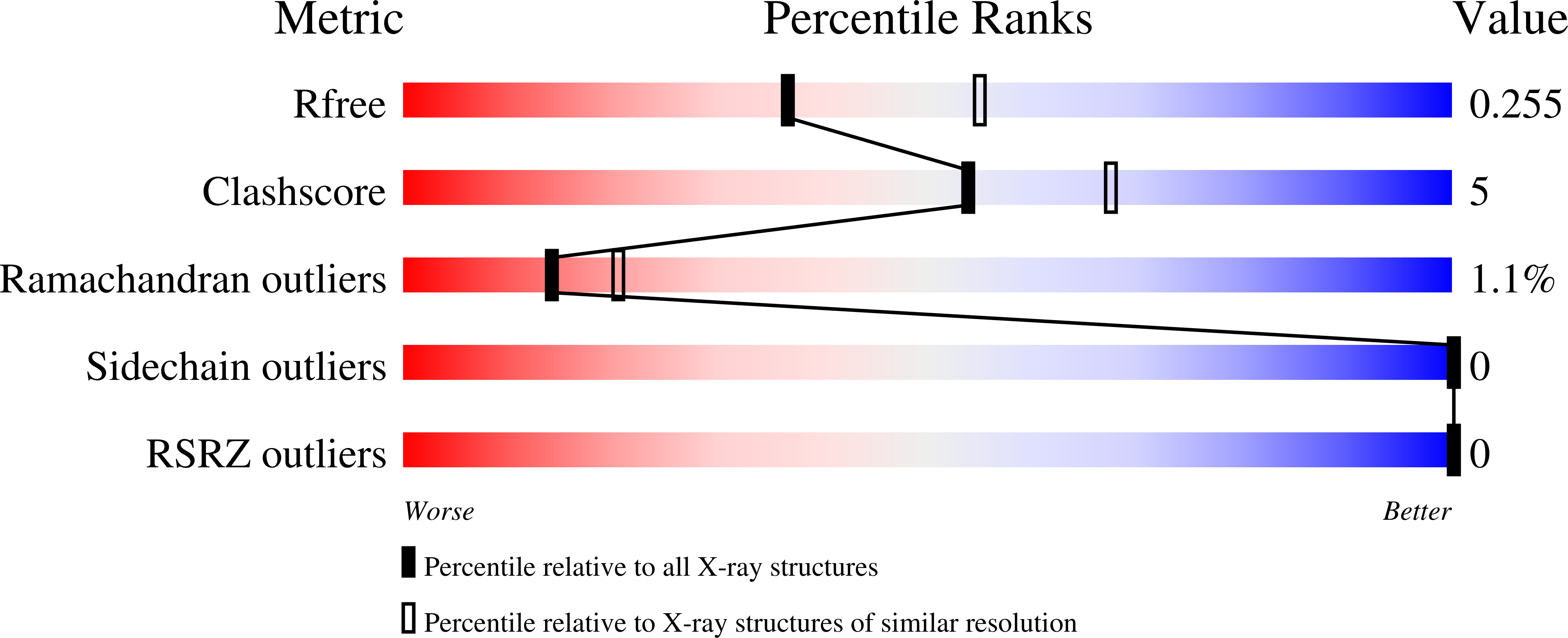
6WKO image
Deposition Date 2020-04-16
Release Date 2021-05-19
Last Version Date 2023-10-18
Entry Detail
PDB ID:
6WKO
Keywords:
Title:
Structure of an influenza C virus hemagglutinin-esterase-fusion (HEF2) intermediate
Biological Source:
Method Details:
Experimental Method:
Resolution:
2.40 Å
R-Value Free:
0.25
R-Value Work:
0.19
R-Value Observed:
0.20
Space Group:
P 21 3
Macromolecular Entities
Structures with similar UniProt ID
Protein Blast
Polymer Type:polypeptide(L)
Molecule:Hemagglutinin-esterase-fusion glycoprotein
Mutagens:C583S
Chain IDs:A
Chain Length:95
Number of Molecules:1
Biological Source:Influenza C virus (strain C/Johannesburg/1/1966)
Ligand Molecules
Primary Citation
Snapshot of an influenza virus glycoprotein fusion intermediate.
Cell Rep 35 109152 109152 (2021)
PMID: 34010634 DOI: 10.1016/j.celrep.2021.109152

Abstact

Enveloped virus entry requires the fusion of cellular and viral membranes, a process directed by their viral fusion glycoproteins. Our current knowledge of this process has been shaped by structural studies of the pre- and post-fusion conformations of these viral fusogens. These structural snapshots have revealed the start and end states necessary for fusion, but the dynamics of the intermediate conformations have remained unclear. Using the influenza C virus hemagglutinin-esterase-fusion glycoprotein as a model, we report the structural and biophysical characterization of a trapped intermediate. Crystallographic studies revealed a structural reorganization of the C terminus to create a second chain reversal region, resulting in the N and C termini being positioned in opposing directions. Intrinsic tryptophan fluorescence and bimane-induced quenching measurements suggest intermediate formation is mediated by conserved hydrophobic residues. Our study reveals a late-stage extended intermediate structural event. This work adds to our understanding of virus cell fusion.

Legend

Protein

Chemical

Disease

Primary Citation of related structures