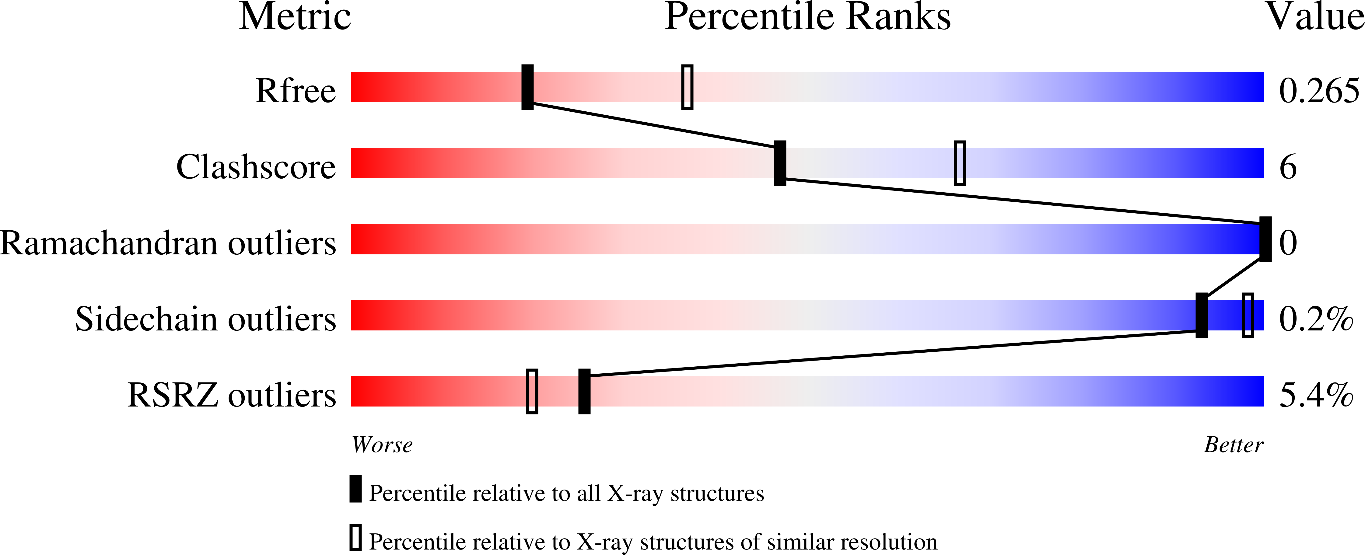
6WIM image
Deposition Date 2020-04-10
Release Date 2020-11-04
Last Version Date 2024-10-09
Entry Detail
PDB ID:
6WIM
Title:
CdiB from Escherichia coli
Biological Source:
Source Organism:
Escherichia coli (Taxon ID: 562)
Method Details:
Experimental Method:
Resolution:
2.60 Å
R-Value Free:
0.26
R-Value Work:
0.24
R-Value Observed:
0.24
Space Group:
P 21 21 2
Macromolecular Entities
Structures with similar UniProt ID
Protein Blast
Polymer Type:polypeptide(L)
Molecule:Outer membrane transporter CdiB
Gene (Uniprot):cdiB
Chain IDs:A
Chain Length:538
Number of Molecules:1
Biological Source:Escherichia coli
Primary Citation
Structural insight into toxin secretion by contact dependent growth inhibition transporters.
Elife 9 ? ? (2020)
PMID: 33089781 DOI: 10.7554/eLife.58100

Abstact

Bacterial contact-dependent growth inhibition (CDI) systems use a type Vb secretion mechanism to export large CdiA toxins across the outer membrane by dedicated outer membrane transporters called CdiB. Here, we report the first crystal structures of two CdiB transporters from Acinetobacter baumannii and Escherichia coli. CdiB transporters adopt a TpsB fold, containing a 16-stranded transmembrane β-barrel connected to two periplasmic domains. The lumen of the CdiB pore is occluded by an N-terminal α-helix and the conserved extracellular loop 6; these two elements adopt different conformations in the structures. We identified a conserved DxxG motif located on strand β1 that connects loop 6 through different networks of interactions. Structural modifications of DxxG induce rearrangement of extracellular loops and alter interactions with the N-terminal α-helix, preparing the system for α-helix ejection. Using structural biology, functional assays, and molecular dynamics simulations, we show how the barrel pore is primed for CdiA toxin secretion.

Legend

Protein

Chemical

Disease

Primary Citation of related structures