6WB6 image
Deposition Date 2020-03-26
Release Date 2020-11-25
Last Version Date 2024-11-13
Entry Detail
PDB ID:
6WB6
Keywords:
Title:
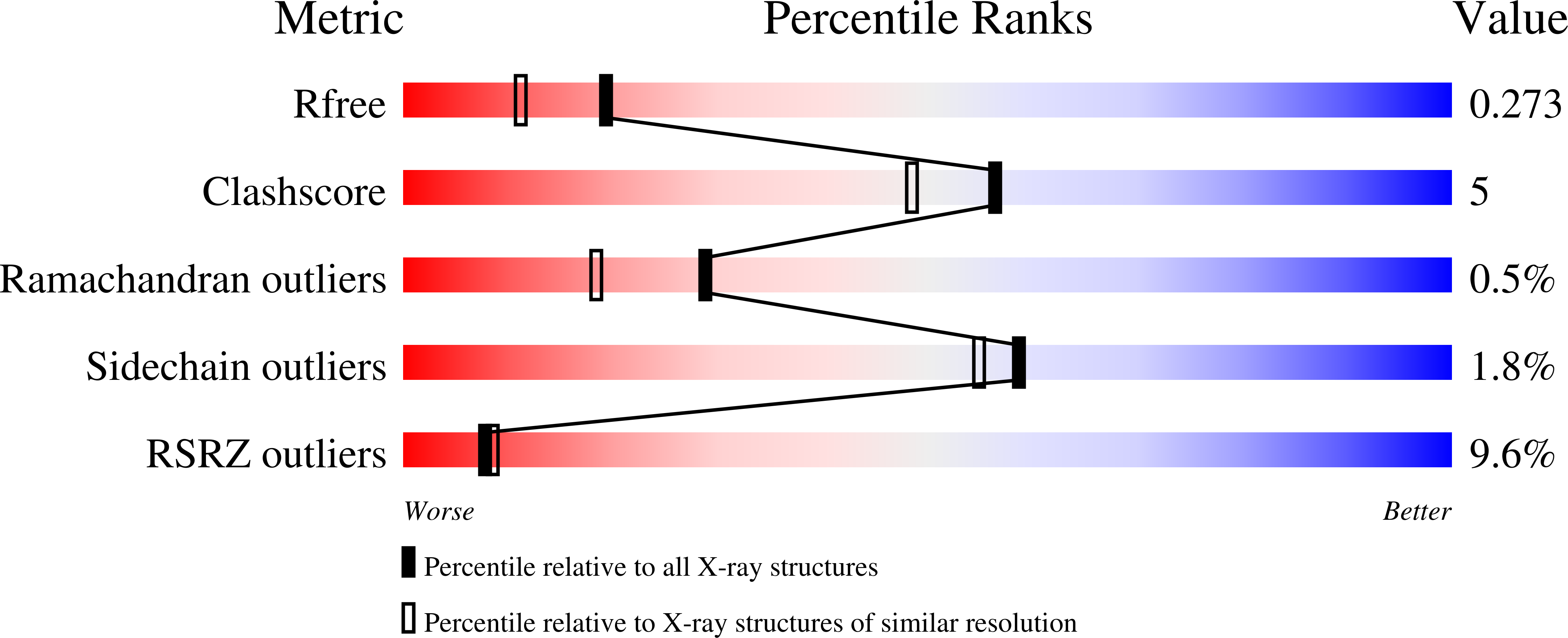
2.05 A resolution structure of transferrin 1 from Manduca sexta
Biological Source:
Source Organism:
Manduca sexta (Taxon ID: 7130)
Method Details:
Experimental Method:
Resolution:
2.05 Å
R-Value Free:
0.27
R-Value Work:
0.20
R-Value Observed:
0.21
Space Group:
P 21 21 21
Macromolecular Entities
Structures with similar UniProt ID
Protein Blast
Polymer Type:polypeptide(L)
Molecule:Transferrin
Chain IDs:A, B
Chain Length:663
Number of Molecules:2
Biological Source:Manduca sexta
Primary Citation
Structural insight into the novel iron-coordination and domain interactions of transferrin-1 from a model insect, Manduca sexta.
Protein Sci. 30 408 422 (2021)
PMID: 33197096 DOI: 10.1002/pro.3999

Abstact

Transferrins function in iron sequestration and iron transport by binding iron tightly and reversibly. Vertebrate transferrins coordinate iron through interactions with two tyrosines, an aspartate, a histidine, and a carbonate anion, and conformational changes that occur upon iron binding and release have been described. Much less is known about the structure and functions of insect transferrin-1 (Tsf1), which is present in hemolymph and influences iron homeostasis mostly by unknown mechanisms. Amino acid sequence and biochemical analyses have suggested that iron coordination by Tsf1 differs from that of the vertebrate transferrins. Here we report the first crystal structure (2.05 Å resolution) of an insect transferrin. Manduca sexta (MsTsf1) in the holo form exhibits a bilobal fold similar to that of vertebrate transferrins, but its carboxyl-lobe adopts a novel orientation and contacts with the amino-lobe. The structure revealed coordination of a single Fe3+ ion in the amino-lobe through Tyr90, Tyr204, and two carbonate anions. One carbonate anion is buried near the ferric ion and is coordinated by four residues, whereas the other carbonate anion is solvent exposed and coordinated by Asn121. Notably, these residues are highly conserved in Tsf1 orthologs. Docking analysis suggested that the solvent exposed carbonate position is capable of binding alternative anions. These findings provide a structural basis for understanding Tsf1 function in iron sequestration and transport in insects as well as insight into the similarities and differences in iron homeostasis between insects and humans.

Legend

Protein

Chemical

Disease

Primary Citation of related structures