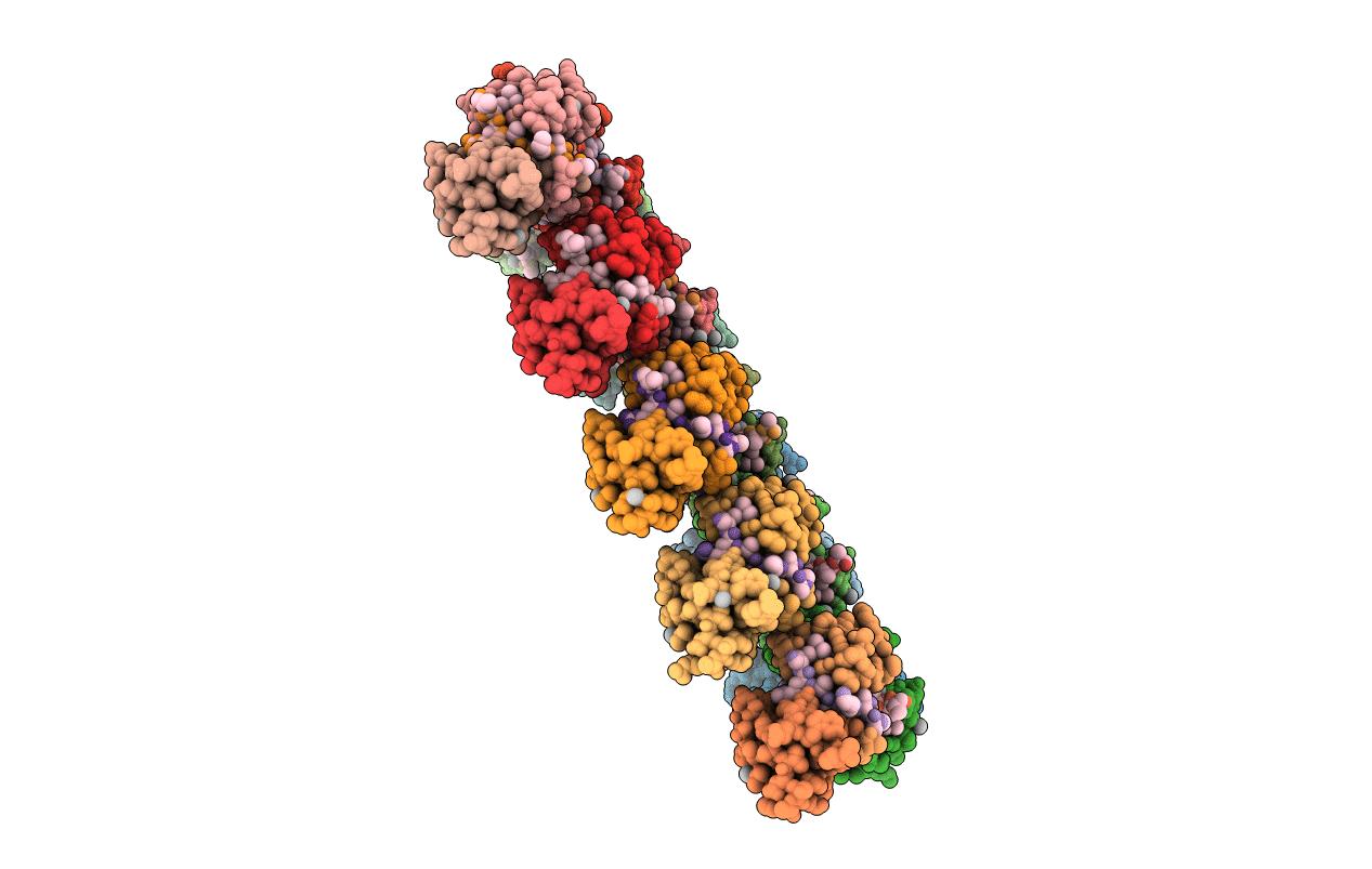
6WAT image
Deposition Date 2020-03-26
Release Date 2020-08-26
Last Version Date 2023-10-18
Entry Detail
PDB ID:
6WAT
Keywords:
Title:
complex structure of PHF1
Biological Source:
Source Organism:
Homo sapiens (Taxon ID: 9606)
Method Details:
Experimental Method:
Resolution:
1.80 Å
R-Value Free:
0.26
R-Value Work:
0.23
R-Value Observed:
0.23
Space Group:
P 1 21 1
Macromolecular Entities
Polymer Type:polypeptide(L)
Molecule:Histone H3.1t peptide
Gene (Uniprot):H3-4
Chain IDs:EA (auth: A), FA (auth: B), GA (auth: C), HA (auth: D), IA (auth: E), JA (auth: F), KA (auth: G), LA (auth: H), MA (auth: I), NA (auth: J), OA (auth: K), PA (auth: L), QA (auth: M), RA (auth: N), SA (auth: O), TA (auth: P), UA (auth: Q), VA (auth: R), WA (auth: S), XA (auth: T), YA (auth: V), ZA (auth: Y), AB (auth: Z), BB (auth: a), CB (auth: b), DB (auth: c), EB (auth: d), FB (auth: e), GB (auth: f), HB (auth: g)
Chain Length:9
Number of Molecules:30
Biological Source:Homo sapiens
Polymer Type:polypeptide(L)
Molecule:PHD finger protein 1
Gene (Uniprot):PHF1
Chain IDs:A (auth: AA), B (auth: AC), C (auth: BA), D (auth: BC), E (auth: CA), F (auth: CC), G (auth: DA), H (auth: DC), I (auth: EA), J (auth: EC), K (auth: FA), L (auth: FC), M (auth: GA), N (auth: GC), O (auth: HA), P (auth: HC), Q (auth: IA), R (auth: IC), S (auth: JA), T (auth: JC), U (auth: KA), V (auth: KC), W (auth: LA), X (auth: LC), Y (auth: MA), Z (auth: MC), AA (auth: NA), BA (auth: NC), CA (auth: OA), DA (auth: OC)
Chain Length:61
Number of Molecules:30
Biological Source:Homo sapiens
Modified Residue
Compound ID Chain ID Parent Comp ID Details 2D Image
AYA EA ALA modified residue
M3L EA LYS modified residue
Ligand Molecules
Primary Citation
Structural basis for histone variant H3tK27me3 recognition by PHF1 and PHF19.
Elife 9 ? ? (2020)
PMID: 32869745 DOI: 10.7554/eLife.58675

Abstact

The Polycomb repressive complex 2 (PRC2) is a multicomponent histone H3K27 methyltransferase complex, best known for silencing the Hox genes during embryonic development. The Polycomb-like proteins PHF1, MTF2, and PHF19 are critical components of PRC2 by stimulating its catalytic activity in embryonic stem cells. The Tudor domains of PHF1/19 have been previously shown to be readers of H3K36me3 in vitro. However, some other studies suggest that PHF1 and PHF19 co-localize with the H3K27me3 mark but not H3K36me3 in cells. Here, we provide further evidence that PHF1 co-localizes with H3t in testis and its Tudor domain preferentially binds to H3tK27me3 over canonical H3K27me3 in vitro. Our complex structures of the Tudor domains of PHF1 and PHF19 with H3tK27me3 shed light on the molecular basis for preferential recognition of H3tK27me3 by PHF1 and PHF19 over canonical H3K27me3, implicating that H3tK27me3 might be a physiological ligand of PHF1/19.

Legend

Protein

Chemical

Disease

Primary Citation of related structures