Deposition Date
2020-03-13
Release Date
2020-05-06
Last Version Date
2023-10-18
Entry Detail
PDB ID:
6W5Q
Keywords:
Title:
Structure of the globular C-terminal domain of P. aeruginosa LpoP
Biological Source:
Source Organism(s):
Pseudomonas aeruginosa (Taxon ID: 287)
Expression System(s):
Method Details:
Experimental Method:
Resolution:
2.20 Å
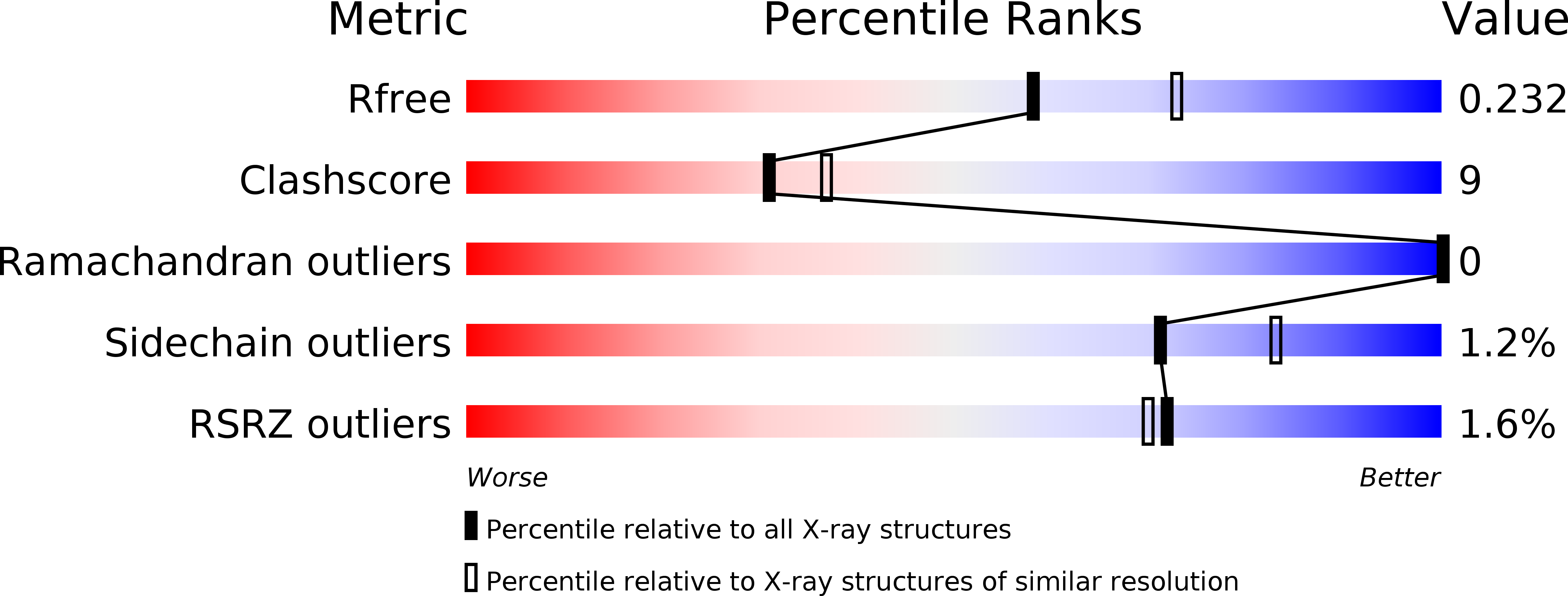
R-Value Free:
0.22
R-Value Work:
0.19
R-Value Observed:
0.19
Space Group:
P 1 21 1