6VXK image
Deposition Date 2020-02-22
Release Date 2020-04-29
Last Version Date 2024-11-13
Entry Detail
PDB ID:
6VXK
Title:
Cryo-EM Structure of the full-length A39R/PlexinC1 complex
Biological Source:
Source Organism:
Host Organism:
Method Details:
Experimental Method:
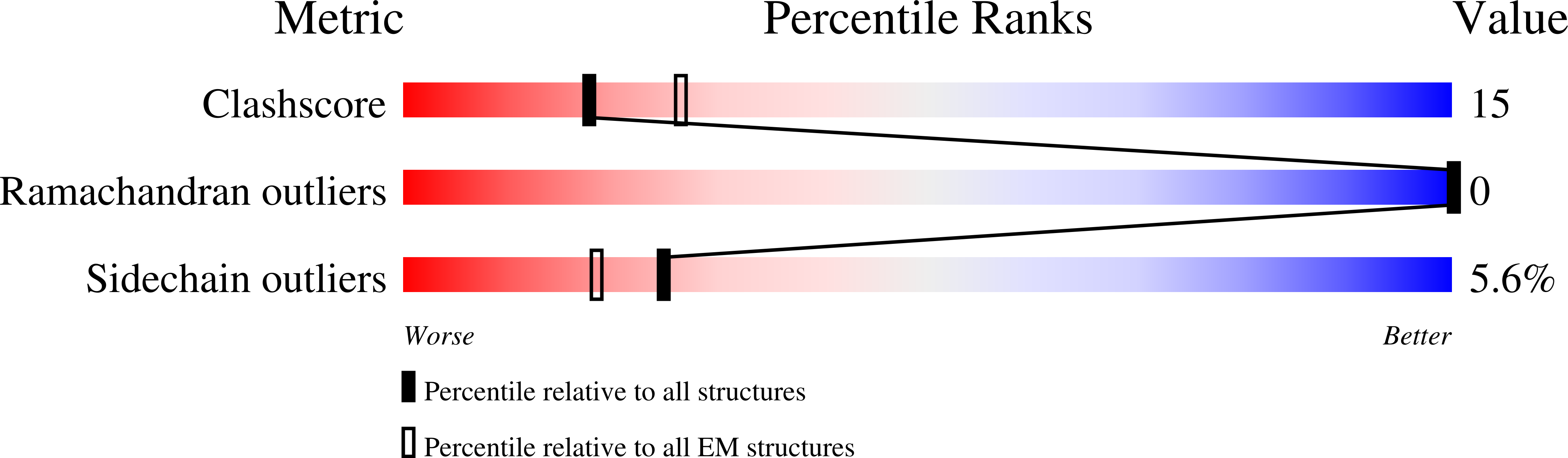
Resolution:
3.10 Å
Aggregation State:
PARTICLE
Reconstruction Method:
SINGLE PARTICLE
Macromolecular Entities
Structures with similar UniProt ID
Protein Blast
Polymer Type:polypeptide(L)
Molecule:Semaphorin-like protein 139
Gene (Uniprot):EVM139
Chain IDs:A, C
Chain Length:398
Number of Molecules:2
Biological Source:Ectromelia virus (strain Moscow)
Structures with similar UniProt ID
Protein Blast
Polymer Type:polypeptide(L)
Molecule:Plexin-C1
Gene (Uniprot):PLXNC1
Chain IDs:B, D
Chain Length:1545
Number of Molecules:2
Biological Source:Homo sapiens
Ligand Molecules
Primary Citation
Cryo-EM structure of the PlexinC1/A39R complex reveals inter-domain interactions critical for ligand-induced activation.
Nat Commun 11 1953 1953 (2020)
PMID: 32327662 DOI: 10.1038/s41467-020-15862-0

Abstact

Plexins are receptors for semaphorins that transduce signals for regulating neuronal development and other processes. Plexins are single-pass transmembrane proteins with multiple domains in both the extracellular and intracellular regions. Semaphorin activates plexin by binding to its extracellular N-terminal Sema domain, inducing the active dimer of the plexin intracellular region. The mechanism underlying this activation process of plexin is incompletely understood. We present cryo-electron microscopic structure of full-length human PlexinC1 in complex with the viral semaphorin mimic A39R. The structure shows that A39R induces a specific dimer of PlexinC1 where the membrane-proximal domains from the two PlexinC1 protomers are placed close to each other, poised to promote the active dimer of the intracellular region. This configuration is imposed by a distinct conformation of the PlexinC1 extracellular region, stabilized by inter-domain interactions among the Sema and membrane-proximal domains. Our mutational analyses support the critical role of this conformation in PlexinC1 activation.

Legend

Protein

Chemical

Disease

Primary Citation of related structures