6VW7 image
Deposition Date 2020-02-18
Release Date 2020-04-08
Last Version Date 2024-03-06
Entry Detail
PDB ID:
6VW7
Keywords:
Title:
Formate Dehydrogenase FdsABG subcomplex FdsBG from C. necator - NADH bound
Biological Source:
Source Organism:
Host Organism:
Method Details:
Experimental Method:
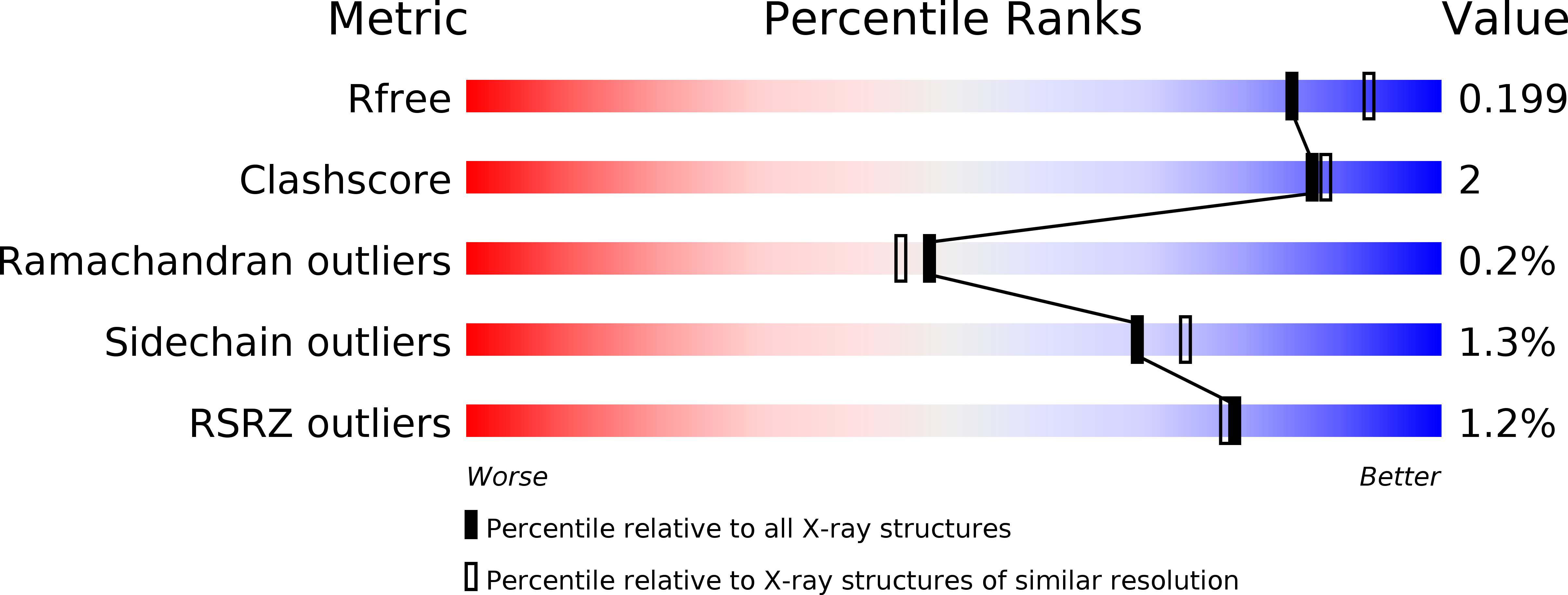
Resolution:
2.00 Å
R-Value Free:
0.19
R-Value Work:
0.15
R-Value Observed:
0.15
Space Group:
C 1 2 1
Macromolecular Entities
Structures with similar UniProt ID
Protein Blast
Polymer Type:polypeptide(L)
Molecule:NAD-dependent formate dehydrogenase gamma subunit
Gene (Uniprot):fdsG
Chain IDs:A, C
Chain Length:176
Number of Molecules:2
Biological Source:Cupriavidus necator
Structures with similar UniProt ID
Protein Blast
Polymer Type:polypeptide(L)
Molecule:NAD-dependent formate dehydrogenase beta subunit
Gene (Uniprot):fdsB
Chain IDs:B, D
Chain Length:520
Number of Molecules:2
Biological Source:Cupriavidus necator
Primary Citation
Crystallographic and kinetic analyses of the FdsBG subcomplex of the cytosolic formate dehydrogenase FdsABG fromCupriavidus necator.
J.Biol.Chem. 295 6570 6585 (2020)
PMID: 32249211 DOI: 10.1074/jbc.RA120.013264

Abstact

Formate oxidation to carbon dioxide is a key reaction in one-carbon compound metabolism, and its reverse reaction represents the first step in carbon assimilation in the acetogenic and methanogenic branches of many anaerobic organisms. The molybdenum-containing dehydrogenase FdsABG is a soluble NAD+-dependent formate dehydrogenase and a member of the NADH dehydrogenase superfamily. Here, we present the first structure of the FdsBG subcomplex of the cytosolic FdsABG formate dehydrogenase from the hydrogen-oxidizing bacterium Cupriavidus necator H16 both with and without bound NADH. The structures revealed that the two iron-sulfur clusters, Fe4S4 in FdsB and Fe2S2 in FdsG, are closer to the FMN than they are in other NADH dehydrogenases. Rapid kinetic studies and EPR measurements of rapid freeze-quenched samples of the NADH reduction of FdsBG identified a neutral flavin semiquinone, FMNH•, not previously observed to participate in NADH-mediated reduction of the FdsABG holoenzyme. We found that this semiquinone forms through the transfer of one electron from the fully reduced FMNH-, initially formed via NADH-mediated reduction, to the Fe2S2 cluster. This Fe2S2 cluster is not part of the on-path chain of iron-sulfur clusters connecting the FMN of FdsB with the active-site molybdenum center of FdsA. According to the NADH-bound structure, the nicotinamide ring stacks onto the re-face of the FMN. However, NADH binding significantly reduced the electron density for the isoalloxazine ring of FMN and induced a conformational change in residues of the FMN-binding pocket that display peptide-bond flipping upon NAD+ binding in proper NADH dehydrogenases.

Legend

Protein

Chemical

Disease

Primary Citation of related structures