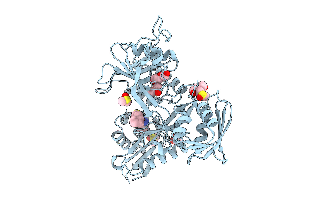
6VUU image
Entry Detail
PDB ID:
6VUU
Title:
Crystal structure of Eis from Mycobacterium tuberculosis in complex with inhibitor SGT1347
Biological Source:
PDB Version:
Deposition Date:
2020-02-16
Release Date:
2020-06-03
Method Details:
Experimental Method:
Resolution:
2.60 Å
R-Value Free:
0.22
R-Value Work:
0.17
R-Value Observed:
0.17
Space Group:
H 3 2
Macromolecular Entities
Polymer Type:polypeptide(L)
Description:N-acetyltransferase Eis
Mutations:C204A
Chain IDs:A
Chain Length:422
Number of Molecules:1
Biological Source:Mycobacterium tuberculosis (strain ATCC 25618 / H37Rv)
Primary Citation
Structure-Guided Optimization of Inhibitors of Acetyltransferase Eis fromMycobacterium tuberculosis.
Acs Chem.Biol. 15 1581 1594 (2020)
PMID: 32421305 DOI: 10.1021/acschembio.0c00184

Abstact

The enhanced intracellular survival (Eis) protein of Mycobacterium tuberculosis (Mtb) is a versatile acetyltransferase that multiacetylates aminoglycoside antibiotics abolishing their binding to the bacterial ribosome. When overexpressed as a result of promoter mutations, Eis causes drug resistance. In an attempt to overcome the Eis-mediated kanamycin resistance of Mtb, we designed and optimized structurally unique thieno[2,3-d]pyrimidine Eis inhibitors toward effective kanamycin adjuvant combination therapy. We obtained 12 crystal structures of enzyme-inhibitor complexes, which guided our rational structure-based design of 72 thieno[2,3-d]pyrimidine analogues divided into three families. We evaluated the potency of these inhibitors in vitro as well as their ability to restore the activity of kanamycin in a resistant strain of Mtb, in which Eis was upregulated. Furthermore, we evaluated the metabolic stability of 11 compounds in vitro. This study showcases how structural information can guide Eis inhibitor design.

Legend

Protein

Chemical

Disease

Primary Citation of related structures