Deposition Date
2020-02-04
Release Date
2020-12-02
Last Version Date
2023-10-11
Entry Detail
PDB ID:
6VPT
Keywords:
Title:
Crystal structure and mechanistic molecular modeling studies of Rv3377c: the Mycobacterium tuberculosis diterpene cyclase
Biological Source:
Source Organism(s):
Mycobacterium tuberculosis (Taxon ID: 1773)
Expression System(s):
Method Details:
Experimental Method:
Resolution:
2.72 Å
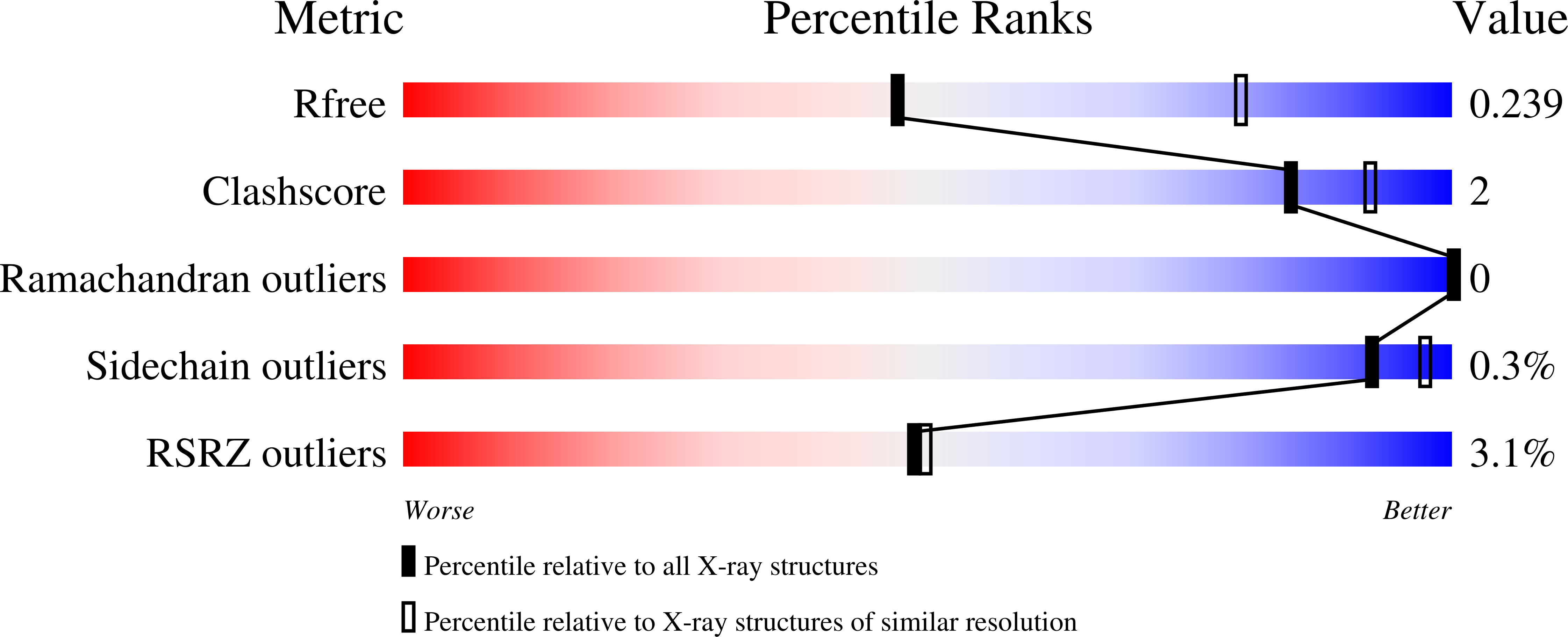
R-Value Free:
0.23
R-Value Work:
0.21
R-Value Observed:
0.21
Space Group:
C 1 2 1