6VLO image
Deposition Date 2020-01-24
Release Date 2020-03-04
Last Version Date 2023-10-11
Entry Detail
PDB ID:
6VLO
Keywords:
Title:
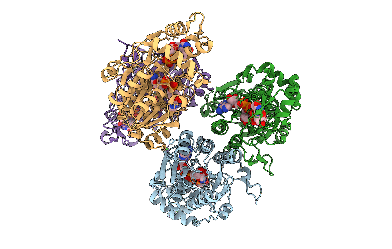
X-ray Structure of the R141 Sugar 4,6-dehydratase from Acanthamoeba polyphaga Minivirus
Biological Source:
Source Organism:
Host Organism:
Method Details:
Experimental Method:
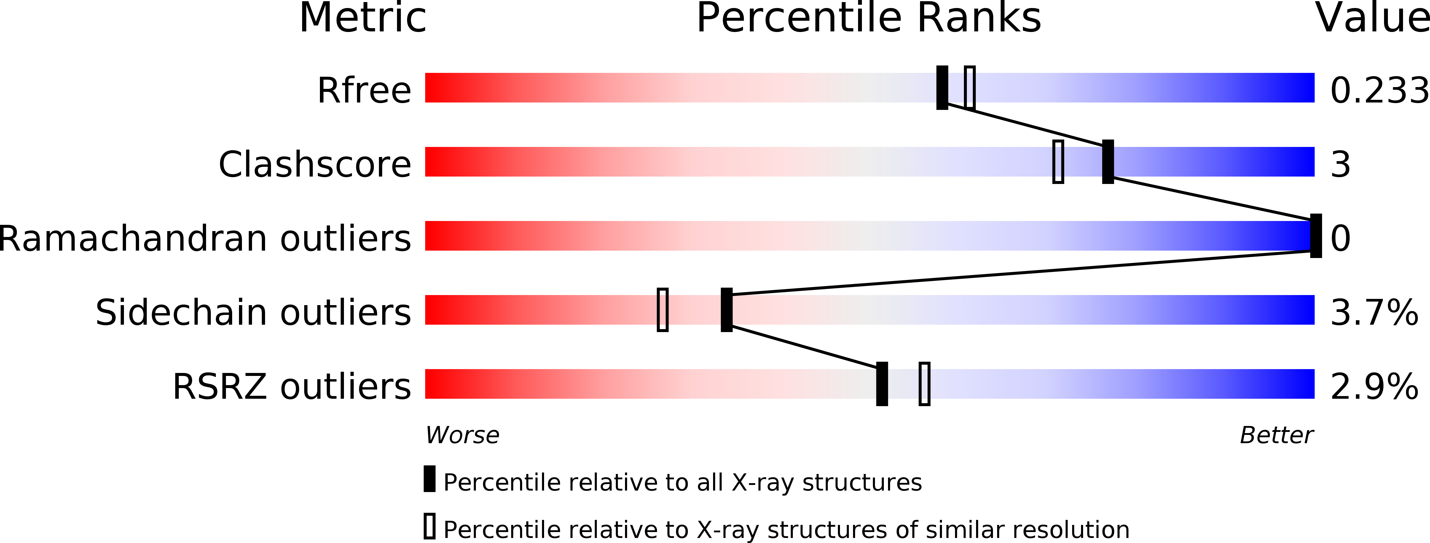
Resolution:
2.05 Å
R-Value Free:
0.22
R-Value Work:
0.18
R-Value Observed:
0.18
Space Group:
P 21 21 21
Macromolecular Entities
Polymer Type:polypeptide(L)
Molecule:Putative dTDP-D-glucose 4,6-dehydratase
Gene (Uniprot):MIMI_R141
Chain IDs:A, B, C, D
Chain Length:326
Number of Molecules:4
Biological Source:Acanthamoeba polyphaga mimivirus
Primary Citation
Biochemical analysis of a sugar 4,6-dehydratase from Acanthamoeba polyphaga Mimivirus.
Protein Sci. 29 1148 1159 (2020)
PMID: 32083779 DOI: 10.1002/pro.3843

Abstact

The exciting discovery of the giant DNA Mimivirus in 2003 challenged the conventional description of viruses in a radical way, and since then, dozens of additional giant viruses have been identified. It has now been demonstrated that the Mimivirus genome encodes for the two enzymes required for the production of the unusual sugar 4-amino-4,6-dideoxy-d-glucose, namely a 4,6-dehydratase and an aminotransferase. In light of our long-standing interest in the bacterial 4,6-dehydratases and in unusual sugars in general, we conducted a combined structural and functional analysis of the Mimivirus 4,6-dehydratase referred to as R141. For this investigation, the three-dimensional X-ray structure of R141 was determined to 2.05 Å resolution and refined to an R-factor of 18.3%. The overall fold of R141 places it into the short-chain dehydrogenase/reductase (SDR) superfamily of proteins. Whereas its molecular architecture is similar to that observed for the bacterial 4,6-dehydratases, there are two key regions where the polypeptide chain adopts different conformations. In particular, the conserved tyrosine that has been implicated as a catalytic acid or base in SDR superfamily members is splayed away from the active site by nearly 12 Å, thereby suggesting that a major conformational change must occur upon substrate binding. In addition to the structural analysis, the kinetic parameters for R141 using either dTDP-d-glucose or UDP-d-glucose as substrates were determined. Contrary to a previous report, R141 demonstrates nearly identical catalytic efficiency with either nucleotide-linked sugar. The data presented herein represent the first three-dimensional model for a viral 4,6-dehydratase and thus expands our understanding of these fascinating enzymes.

Legend

Protein

Chemical

Disease

Primary Citation of related structures