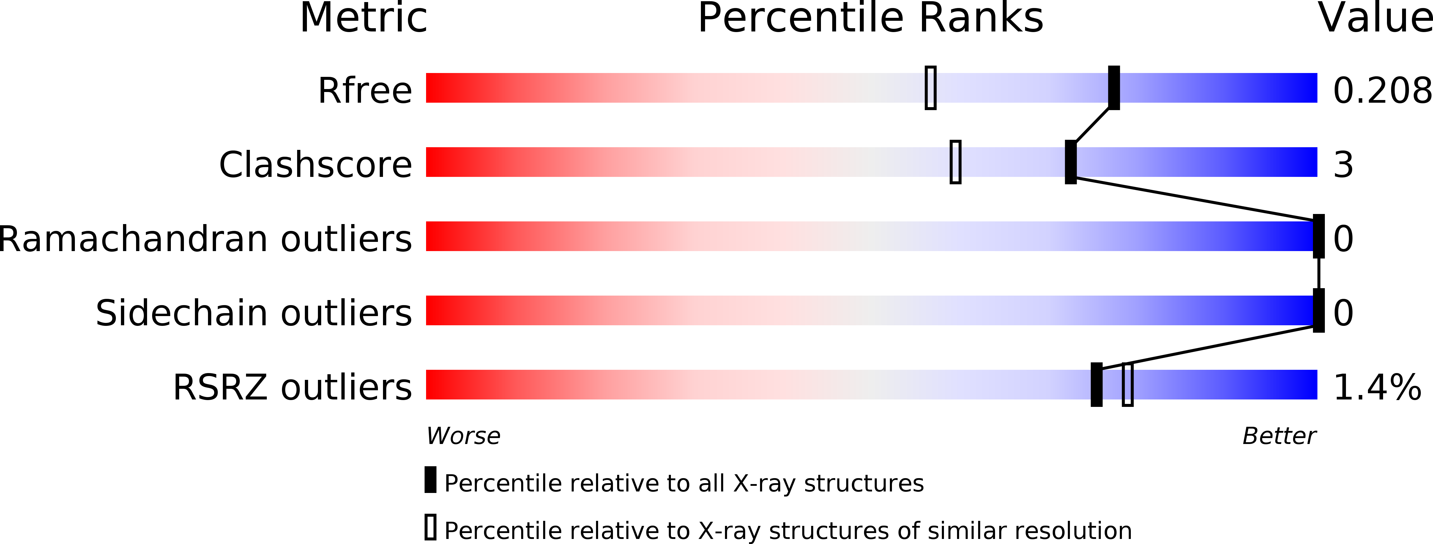
6VGV image
Deposition Date 2020-01-09
Release Date 2020-05-27
Last Version Date 2024-04-03
Entry Detail
PDB ID:
6VGV
Keywords:
Title:
Crystal structure of VidaL intein
Biological Source:
Source Organism:
Host Organism:
Method Details:
Experimental Method:
Resolution:
1.65 Å
R-Value Free:
0.20
R-Value Work:
0.16
R-Value Observed:
0.16
Space Group:
C 1 2 1
Macromolecular Entities
Polymer Type:polypeptide(L)
Molecule:VidaL
Mutagens:C4A, N142A
Chain IDs:A
Chain Length:145
Number of Molecules:1
Biological Source:synthetic construct
Ligand Molecules
Primary Citation
Live-cell protein engineering with an ultra-short split intein.
Proc.Natl.Acad.Sci.USA 117 12041 12049 (2020)
PMID: 32424098 DOI: 10.1073/pnas.2003613117

Abstact

Split inteins are privileged molecular scaffolds for the chemical modification of proteins. Though efficient for in vitro applications, these polypeptide ligases have not been utilized for the semisynthesis of proteins in live cells. Here, we biochemically and structurally characterize the naturally split intein VidaL. We show that this split intein, which features the shortest known N-terminal fragment, supports rapid and efficient protein trans-splicing under a range of conditions, enabling semisynthesis of modified proteins both in vitro and in mammalian cells. The utility of this protein engineering system is illustrated through the traceless assembly of multidomain proteins whose biophysical properties render them incompatible with a single expression system, as well as by the semisynthesis of dual posttranslationally modified histone proteins in live cells. We also exploit the domain swapping function of VidaL to effect simultaneous modification and translocation of the nuclear protein HP1α in live cells. Collectively, our studies highlight the VidaL system as a tool for the precise chemical modification of cellular proteins with spatial and temporal control.

Legend

Protein

Chemical

Disease

Primary Citation of related structures