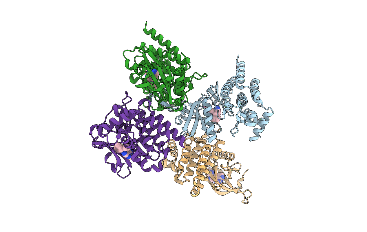
6VGL image
Deposition Date 2020-01-08
Release Date 2021-01-13
Last Version Date 2024-11-06
Entry Detail
PDB ID:
6VGL
Title:
JAK2 JH1 in complex with ruxolitinib
Biological Source:
Source Organism:
Homo sapiens (Taxon ID: 9606)
Host Organism:
Method Details:
Experimental Method:
Resolution:
1.90 Å
R-Value Free:
0.21
R-Value Work:
0.18
R-Value Observed:
0.18
Space Group:
P 1 21 1
Macromolecular Entities
Polymer Type:polypeptide(L)
Molecule:Tyrosine-protein kinase JAK2
Gene (Uniprot):JAK2
Chain IDs:A (auth: D), B, C, D (auth: A)
Chain Length:308
Number of Molecules:4
Biological Source:Homo sapiens
Modified Residue
Compound ID Chain ID Parent Comp ID Details 2D Image
PTR A TYR modified residue
Ligand Molecules
Primary Citation
Structural Insights into JAK2 Inhibition by Ruxolitinib, Fedratinib, and Derivatives Thereof.
J.Med.Chem. 64 2228 2241 (2021)
PMID: 33570945 DOI: 10.1021/acs.jmedchem.0c01952

Abstact

The discovery that aberrant activity of Janus kinase 2 (JAK2) is a driver of myeloproliferative neoplasms (MPNs) has led to significant efforts to develop small molecule inhibitors for this patient population. Ruxolitinib and fedratinib have been approved for use in MPN patients, while baricitinib, an achiral analogue of ruxolitinib, has been approved for rheumatoid arthritis. However, structural information on the interaction of these therapeutics with JAK2 remains unknown. Here, we describe a new methodology for the large-scale production of JAK2 from mammalian cells, which enabled us to determine the first crystal structures of JAK2 bound to these drugs and derivatives thereof. Along with biochemical and cellular data, the results provide a comprehensive view of the shape complementarity required for chiral and achiral inhibitors to achieve highest activity, which may facilitate the development of more effective JAK2 inhibitors as therapeutics.

Legend

Protein

Chemical

Disease

Primary Citation of related structures