
Deposition Date
2019-12-28
Release Date
2020-11-25
Last Version Date
2023-10-11
Entry Detail
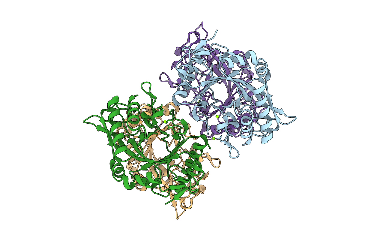
PDB ID:
6VE1
Keywords:
Title:
Crystal structure of endo-beta-N-acetylglucosaminidase H at high pH
Biological Source:
Source Organism(s):
Streptomyces plicatus (Taxon ID: 1922)
Expression System(s):
Method Details:
Experimental Method:
Resolution:
2.10 Å
R-Value Free:
0.25
R-Value Work:
0.22
R-Value Observed:
0.22
Space Group:
P 21 2 21
