6VDG image
Deposition Date 2019-12-27
Release Date 2020-01-15
Last Version Date 2023-10-11
Entry Detail
PDB ID:
6VDG
Keywords:
Title:
Crystal Structure of the Y182A HisF Mutant from Thermotoga maritima
Biological Source:
Source Organism:
Host Organism:
Method Details:
Experimental Method:
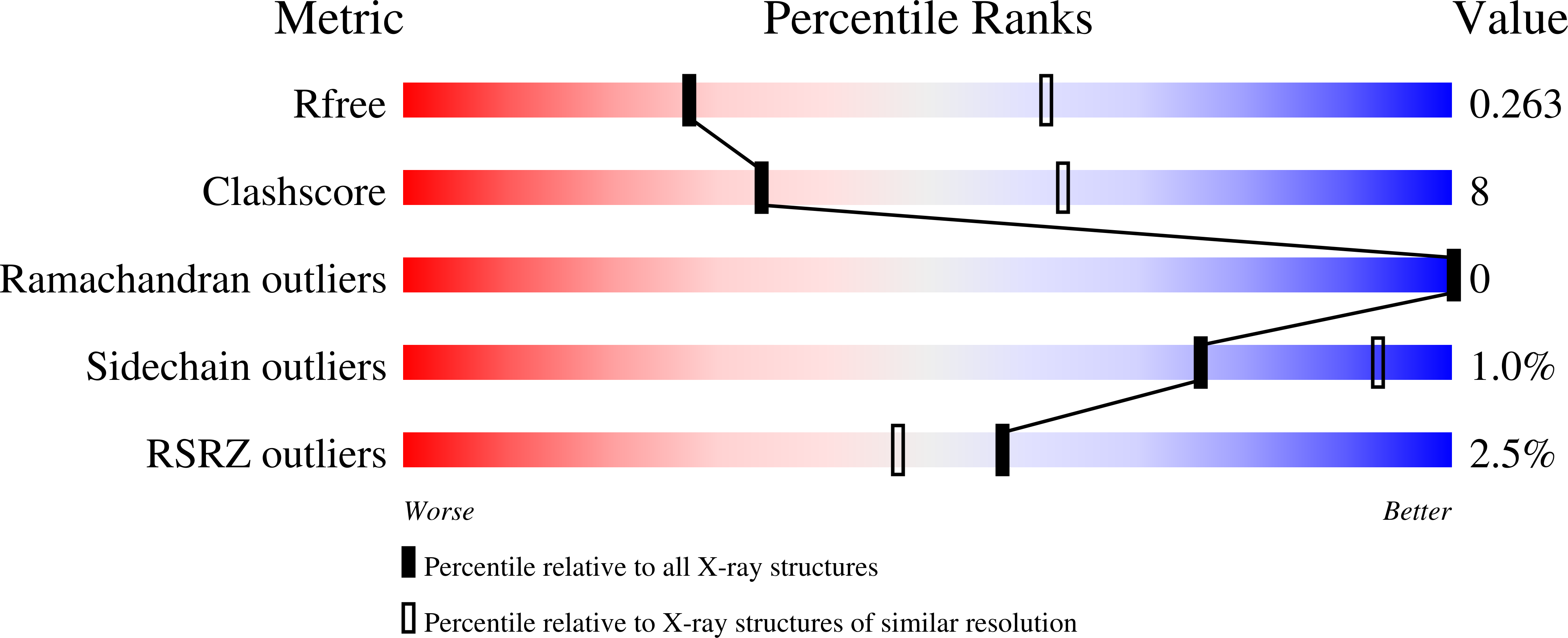
Resolution:
2.79 Å
R-Value Free:
0.26
R-Value Work:
0.20
R-Value Observed:
0.21
Space Group:
P 61 2 2
Macromolecular Entities
Structures with similar UniProt ID
Protein Blast
Polymer Type:polypeptide(L)
Molecule:Imidazole glycerol phosphate synthase subunit HisF
Gene (Uniprot):hisF
Mutagens:Y182A
Chain IDs:A
Chain Length:253
Number of Molecules:1
Biological Source:Thermotoga maritima
Ligand Molecules
Primary Citation
Role of a high centrality residue in protein dynamics and thermal stability.
J.Struct.Biol. 213 107773 107773 (2021)
PMID: 34320379 DOI: 10.1016/j.jsb.2021.107773

Abstact

Centralities determined from Residue Interaction Networks (RIN) in proteins have been used to predict aspects of their structure and dynamics. Here, we correlate the Eigenvector Centrality (Ec) with the rate constant for thermal denaturation (kden) of the HisF protein from Thermotoga maritima based on 12 single alanine substitution mutants. The molecular basis for this correlation was further explored by studying a mutant containing a replacement of a high Ec residue, Y182A, which displayed increased kden at 80 °C. The crystallographic structure of this mutant showed few changes, mostly in two flexible loops. The 1H-15N -HSQC showed only subtle changes of cross peak positions for residues located near the mutation site and scattered throughout the structure. However, the comparison of the RIN showed that Y182 is the vertex of a set of high centrality residues that spreads throughout the HisF structure, which is lacking in the mutant. Cross-correlation displacements of Cα calculated from a molecular dynamics simulation at different temperatures showed that the Y182A mutation reduced the correlated movements in the HisF structure above 70 °C. 1H-15N NMR chemical shift covariance using temperature as perturbation were consistent with these results. In conclusion the increase in temperature drives the structure of the mutant HisF-Y182A into a less connected state, richer in non-concerted motions, located predominantly in the C-terminal half of the protein where Y182 is placed. Conversely, wild-type HisF responds to increased temperature as a single unit. Hence the replacement of a high Ec residue alters the distribution of thermal energy through HisF structure.

Legend

Protein

Chemical

Disease

Primary Citation of related structures
Feedback Form
Name
Email
Institute
Feedback