
Deposition Date
2019-12-17
Release Date
2021-01-27
Last Version Date
2023-10-11
Entry Detail
PDB ID:
6VA9
Keywords:
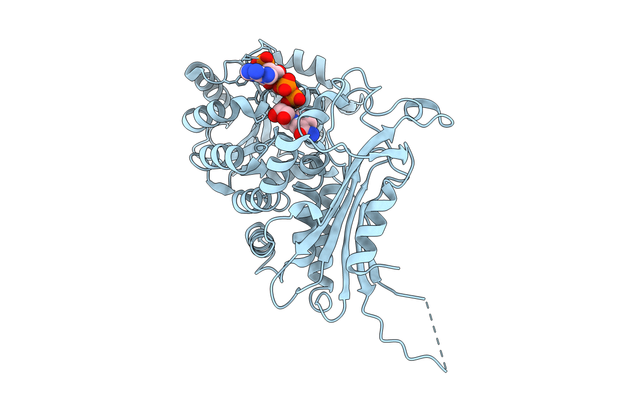
Title:
Crystal structure of glucose-6-phosphate dehydrogenase R393H mutant in complex with catalytic NADP+
Biological Source:
Source Organism(s):
Homo sapiens (Taxon ID: 9606)
Expression System(s):
Method Details:
Experimental Method:
Resolution:
3.95 Å
R-Value Free:
0.22
R-Value Work:
0.19
R-Value Observed:
0.19
Space Group:
P 41 21 2
