Deposition Date
2019-12-16
Release Date
2020-01-22
Last Version Date
2024-03-06
Entry Detail
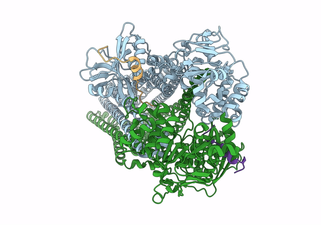
PDB ID:
6V9Z
Keywords:
Title:
Cryo-EM structure of PCAT1 bound to its CtA peptide substrate
Biological Source:
Source Organism(s):
Expression System(s):
Method Details:
Experimental Method:
Resolution:
3.35 Å
Aggregation State:
PARTICLE
Reconstruction Method:
SINGLE PARTICLE