6V4V image
Entry Detail
PDB ID:
6V4V
Title:
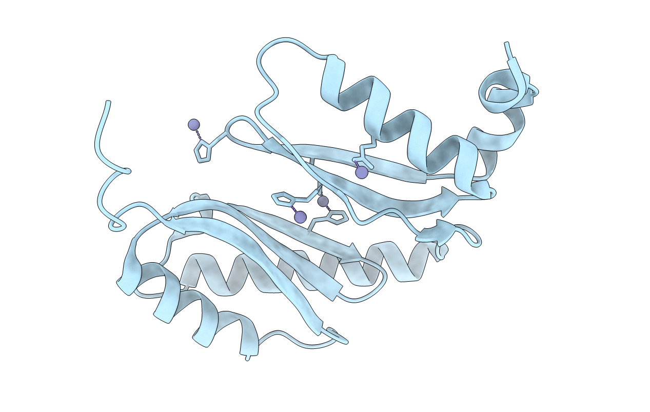
The crystal structure of BonA from Acinetobacter baumannii
Biological Source:
Source Organism:
Host Organism:
PDB Version:
Deposition Date:
2019-12-02
Release Date:
2021-06-02
Method Details:
Experimental Method:
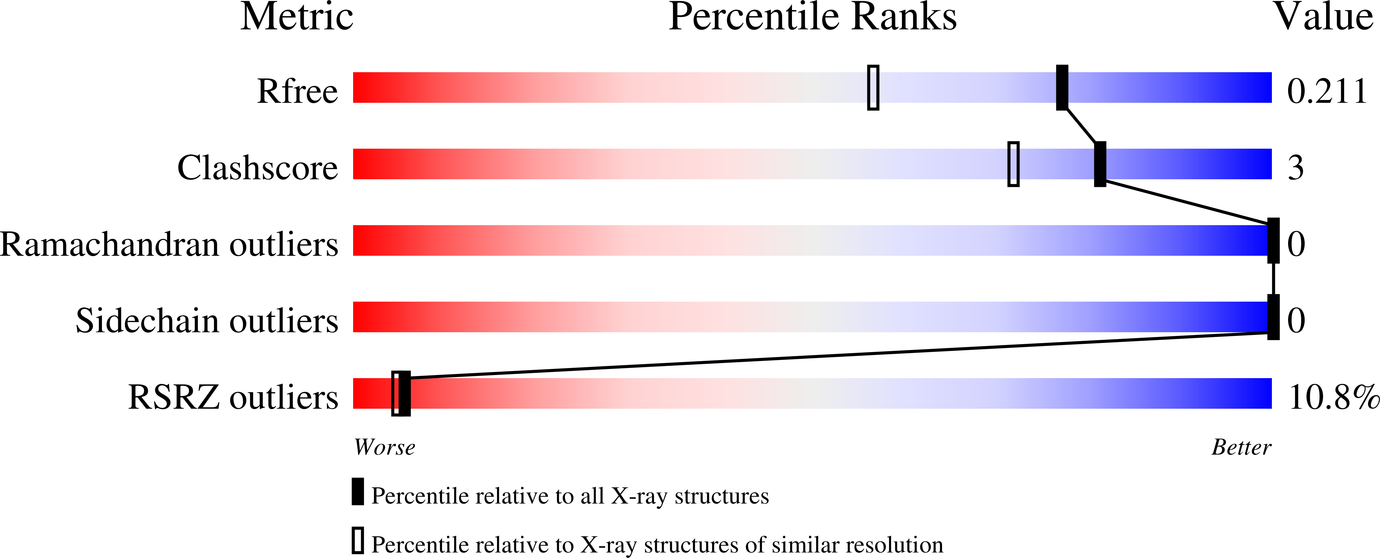
Resolution:
1.65 Å
R-Value Free:
0.21
R-Value Work:
0.18
R-Value Observed:
0.18
Space Group:
P 31 2 1
Macromolecular Entities
Polymer Type:polypeptide(L)
Description:BON domain protein
Chain IDs:A
Chain Length:190
Number of Molecules:1
Biological Source:Acinetobacter baumannii
Modified Residue
Compound ID Chain ID Parent Comp ID Details 2D Image
MSE A MET modified residue
Ligand Molecules
Primary Citation
BonA from Acinetobacter baumannii Forms a Divisome-Localized Decamer That Supports Outer Envelope Function.
Mbio ? e0148021 e0148021 (2021)
PMID: 34311571 DOI: 10.1128/mBio.01480-21

Abstact

Acinetobacter baumannii is a high-risk pathogen due to the rapid global spread of multidrug-resistant lineages. Its phylogenetic divergence from other ESKAPE pathogens means that determinants of its antimicrobial resistance can be difficult to extrapolate from other widely studied bacteria. A recent study showed that A. baumannii upregulates production of an outer membrane lipoprotein, which we designate BonA, in response to challenge with polymyxins. Here, we show that BonA has limited sequence similarity and distinct structural features compared to lipoproteins from other bacterial species. Analyses through X-ray crystallography, small-angle X-ray scattering, electron microscopy, and multiangle light scattering demonstrate that BonA has a dual BON (Bacterial OsmY and Nodulation) domain architecture and forms a decamer via an unusual oligomerization mechanism. This analysis also indicates this decamer is transient, suggesting dynamic oligomerization plays a role in BonA function. Antisera recognizing BonA shows it is an outer membrane protein localized to the divisome. Loss of BonA modulates the density of the outer membrane, consistent with a change in its structure or link to the peptidoglycan, and prevents motility in a clinical strain (ATCC 17978). Consistent with these findings, the dimensions of the BonA decamer are sufficient to permeate the peptidoglycan layer, with the potential to form a membrane-spanning complex during cell division. IMPORTANCE The pathogen Acinetobacter baumannii is considered an urgent threat to human health. A. baumannii is highly resistant to treatment with antibiotics, in part due to its protective cell envelope. This bacterium is only distantly related to other bacterial pathogens, so its cell envelope has distinct properties and contains components distinct from those of other bacteria that support its function. Here, we report the discovery of BonA, a protein that supports A. baumannii outer envelope function and is required for cell motility. We determine the atomic structure of BonA and show that it forms part of the cell division machinery and functions by forming a complex, features that mirror those of distantly related homologs from other bacteria. By improving our understanding of the A. baumannii cell envelope this work will assist in treating this pathogen.

Legend

Protein

Chemical

Disease

Primary Citation of related structures