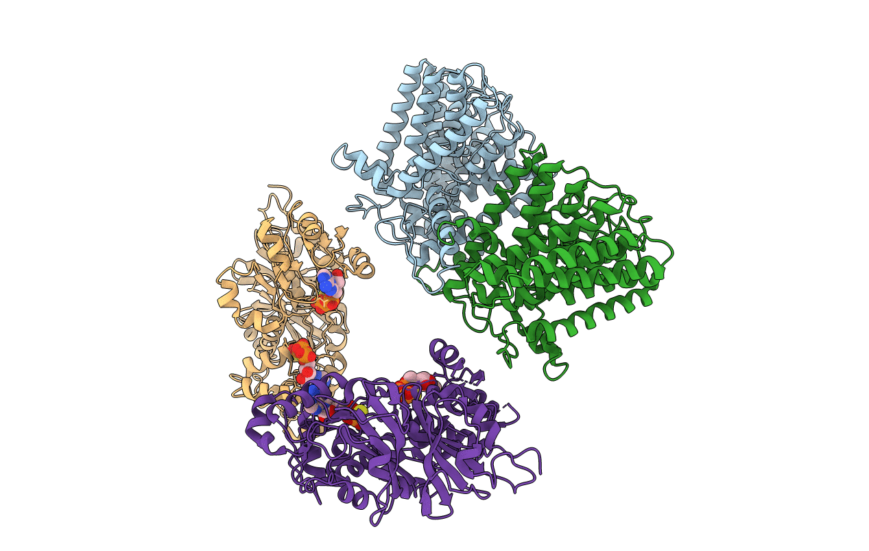
6V4L image
Entry Detail
PDB ID:
6V4L
Title:
Structure of TrkH-TrkA in complex with ATPgammaS
Biological Source:
PDB Version:
Deposition Date:
2019-11-27
Release Date:
2020-02-12
Method Details:
Experimental Method:
Resolution:
3.80 Å
R-Value Free:
0.29
R-Value Work:
0.25
R-Value Observed:
0.25
Space Group:
P 42
Macromolecular Entities
Polymer Type:polypeptide(L)
Description:Trk system potassium uptake protein TrkH
Chain IDs:A, B
Chain Length:485
Number of Molecules:2
Biological Source:Vibrio parahaemolyticus serotype O3:K6 (strain RIMD 2210633)
Polymer Type:polypeptide(L)
Description:Potassium uptake protein TrkA
Chain IDs:C, D
Chain Length:458
Number of Molecules:2
Biological Source:Vibrio parahaemolyticus serotype O3:K6 (strain RIMD 2210633)
Ligand Molecules
Primary Citation
TrkA undergoes a tetramer-to-dimer conversion to open TrkH which enables changes in membrane potential.
Nat Commun 11 547 547 (2020)
PMID: 31992706 DOI: 10.1038/s41467-019-14240-9

Abstact

TrkH is a bacterial ion channel implicated in K+ uptake and pH regulation. TrkH assembles with its regulatory protein, TrkA, which closes the channel when bound to ADP and opens it when bound to ATP. However, it is unknown how nucleotides control the gating of TrkH through TrkA. Here we report the structures of the TrkH-TrkA complex in the presence of ADP or ATP. TrkA forms a tetrameric ring when bound to ADP and constrains TrkH to a closed conformation. The TrkA ring splits into two TrkA dimers in the presence of ATP and releases the constraints on TrkH, resulting in an open channel conformation. Functional studies show that both the tetramer-to-dimer conversion of TrkA and the loss of constraints on TrkH are required for channel gating. In addition, deletion of TrkA in Escherichia coli depolarizes the cell, suggesting that the TrkH-TrkA complex couples changes in intracellular nucleotides to membrane potential.

Legend

Protein

Chemical

Disease

Primary Citation of related structures