Deposition Date
2019-11-25
Release Date
2020-01-08
Last Version Date
2023-10-11
Entry Detail
PDB ID:
6V2T
Keywords:
Title:
X-ray structure of a sugar N-formyltransferase from Shewanella sp FDAARGOS_354
Biological Source:
Source Organism(s):
Shewanella sp. FDAARGOS_354 (Taxon ID: 1930557)
Expression System(s):
Method Details:
Experimental Method:
Resolution:
1.90 Å
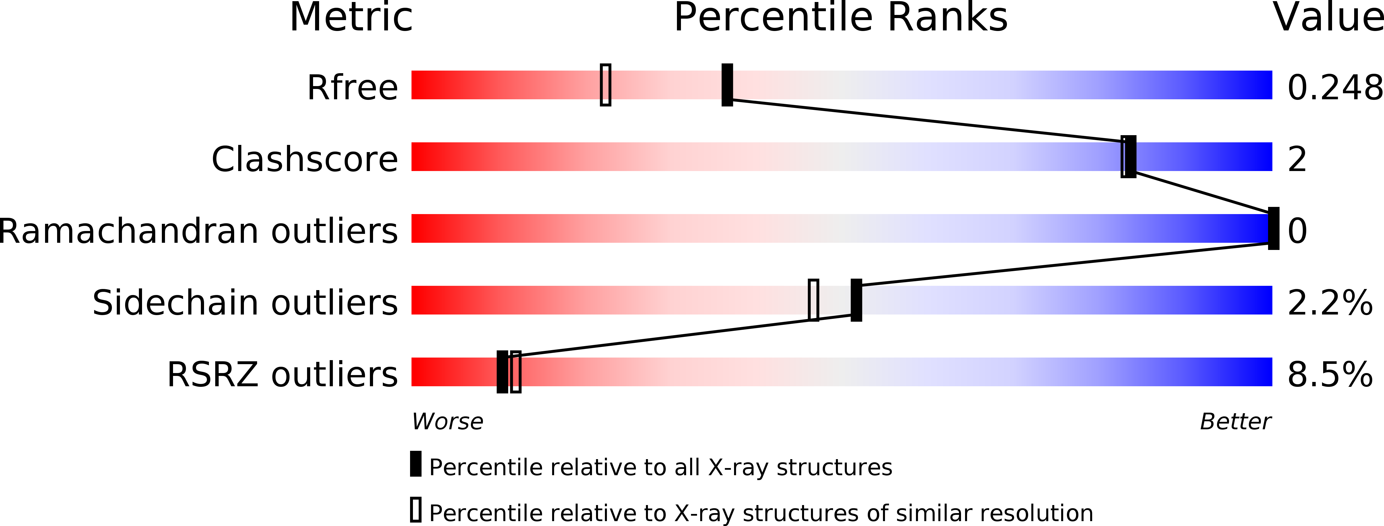
R-Value Free:
0.24
R-Value Work:
0.18
R-Value Observed:
0.18
Space Group:
C 2 2 21