6UZD image
Deposition Date 2019-11-14
Release Date 2020-03-04
Last Version Date 2024-03-06
Entry Detail
PDB ID:
6UZD
Keywords:
Title:
Anthrax toxin protective antigen channels bound to edema factor
Biological Source:
Source Organism:
Method Details:
Experimental Method:
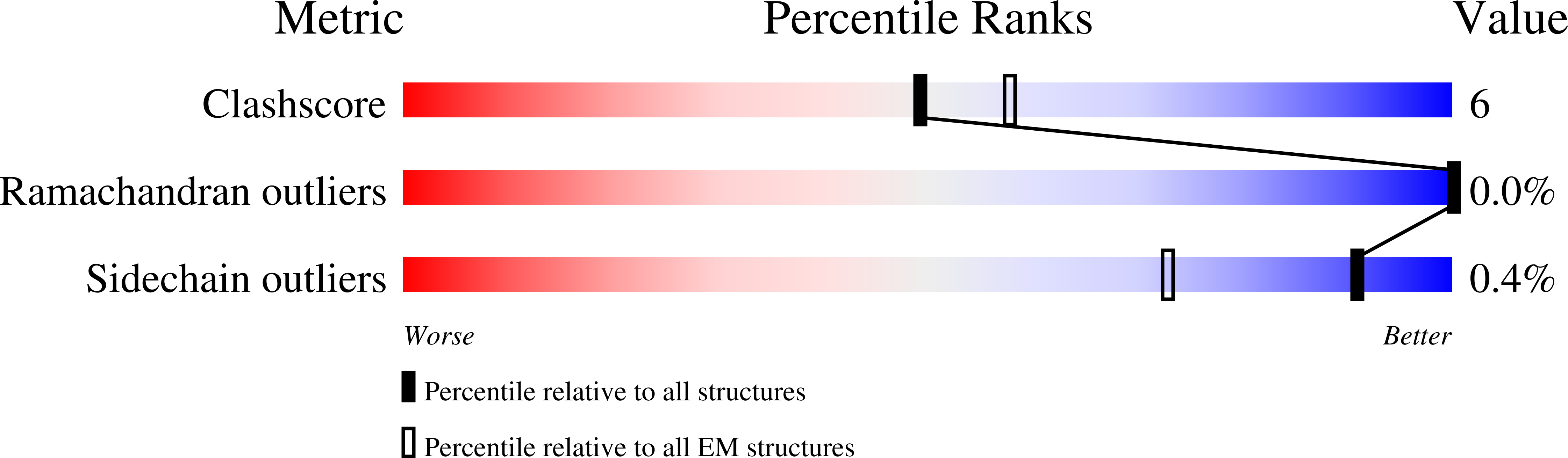
Resolution:
3.40 Å
Aggregation State:
PARTICLE
Reconstruction Method:
SINGLE PARTICLE
Macromolecular Entities
Polymer Type:polypeptide(L)
Molecule:Protective antigen
Gene (Uniprot):pagA
Chain IDs:A, B, C, D, E, F, G
Chain Length:735
Number of Molecules:7
Biological Source:Bacillus anthracis
Polymer Type:polypeptide(L)
Molecule:Calmodulin-sensitive adenylate cyclase
Gene (Uniprot):cya
Chain IDs:H, I
Chain Length:767
Number of Molecules:2
Biological Source:Bacillus anthracis
Ligand Molecules
Primary Citation
Atomic structures of anthrax toxin protective antigen channels bound to partially unfolded lethal and edema factors.
Nat Commun 11 840 840 (2020)
PMID: 32047164 DOI: 10.1038/s41467-020-14658-6

Abstact

Following assembly, the anthrax protective antigen (PA) forms an oligomeric translocon that unfolds and translocates either its lethal factor (LF) or edema factor (EF) into the host cell. Here, we report the cryo-EM structures of heptameric PA channels with partially unfolded LF and EF at 4.6 and 3.1-Å resolution, respectively. The first α helix and β strand of LF and EF unfold and dock into a deep amphipathic cleft, called the α clamp, which resides at the interface of two PA monomers. The α-clamp-helix interactions exhibit structural plasticity when comparing the structures of lethal and edema toxins. EF undergoes a largescale conformational rearrangement when forming the complex with the channel. A critical loop in the PA binding interface is displaced for about 4 Å, leading to the weakening of the binding interface prior to translocation. These structures provide key insights into the molecular mechanisms of translocation-coupled protein unfolding and translocation.

Legend

Protein

Chemical

Disease

Primary Citation of related structures