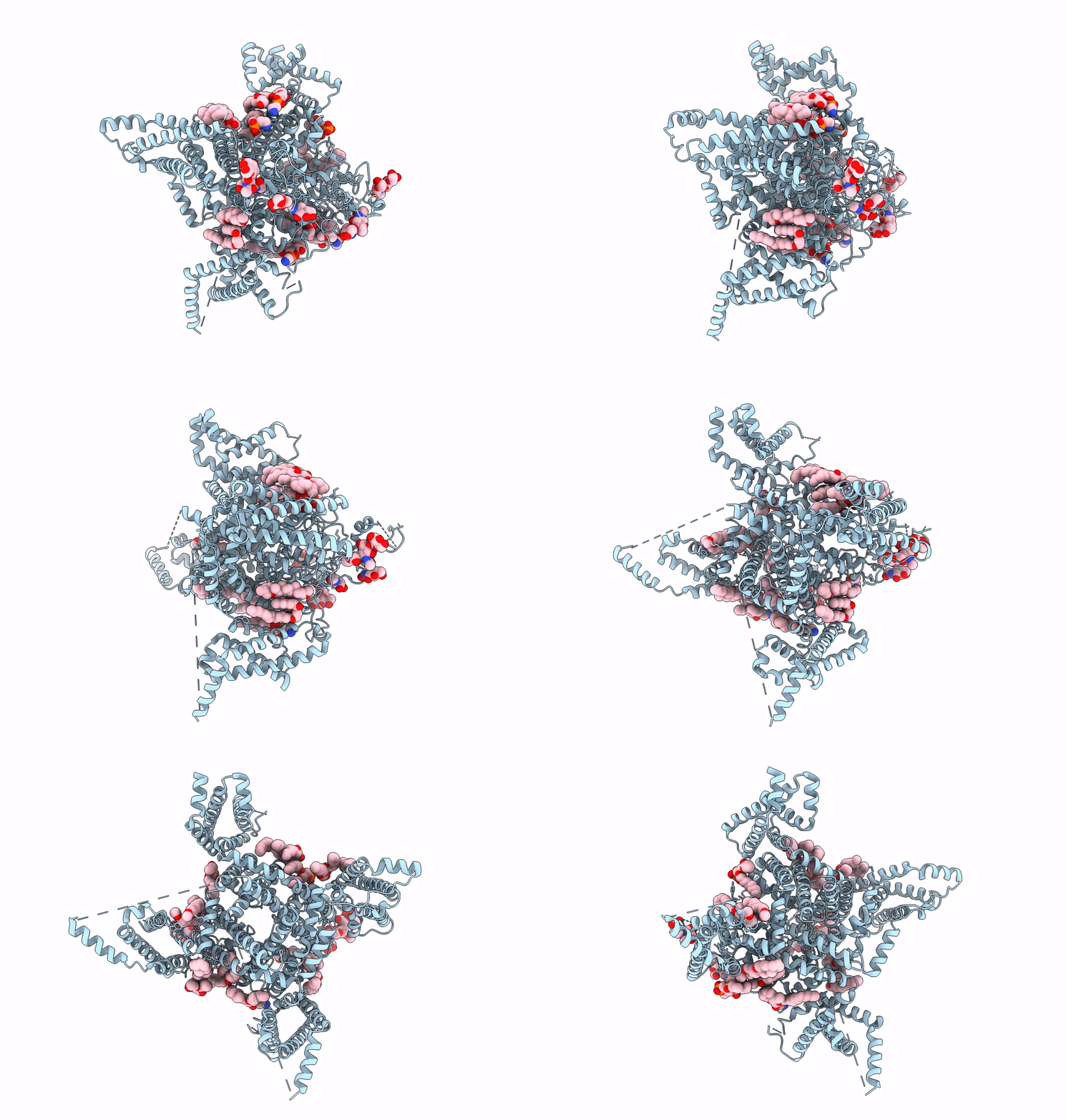
6UZ3 image
Entry Detail
PDB ID:
6UZ3
EMDB ID:
Keywords:
Title:
Cardiac sodium channel
Biological Source:
PDB Version:
Deposition Date:
2019-11-14
Release Date:
2020-01-01
Method Details:
Experimental Method:
Resolution:
3.50 Å
Aggregation State:
PARTICLE
Reconstruction Method:
SINGLE PARTICLE
Macromolecular Entities
Polymer Type:polypeptide(L)
Description:Sodium channel protein type 5 subunit alpha,Green fluorescent protein
Chain IDs:A
Chain Length:1838
Number of Molecules:1
Biological Source:Rattus norvegicus, Aequorea victoria
Primary Citation
Structure of the Cardiac Sodium Channel.
Cell 180 122 134.e10 (2020)
PMID: 31866066 DOI: 10.1016/j.cell.2019.11.041

Abstact

Voltage-gated sodium channel Nav1.5 generates cardiac action potentials and initiates the heartbeat. Here, we report structures of NaV1.5 at 3.2-3.5 Å resolution. NaV1.5 is distinguished from other sodium channels by a unique glycosyl moiety and loss of disulfide-bonding capability at the NaVβ subunit-interaction sites. The antiarrhythmic drug flecainide specifically targets the central cavity of the pore. The voltage sensors are partially activated, and the fast-inactivation gate is partially closed. Activation of the voltage sensor of Domain III allows binding of the isoleucine-phenylalanine-methionine (IFM) motif to the inactivation-gate receptor. Asp and Ala, in the selectivity motif DEKA, line the walls of the ion-selectivity filter, whereas Glu and Lys are in positions to accept and release Na+ ions via a charge-delocalization network. Arrhythmia mutation sites undergo large translocations during gating, providing a potential mechanism for pathogenic effects. Our results provide detailed insights into Nav1.5 structure, pharmacology, activation, inactivation, ion selectivity, and arrhythmias.

Legend

Protein

Chemical

Disease

Primary Citation of related structures